 Movie
Movie Controller


+ Open data

- Basic information
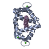
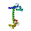
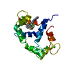
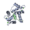
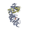
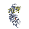
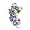
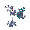
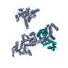
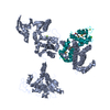
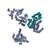
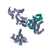
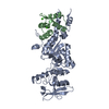
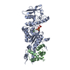
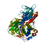
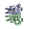
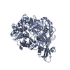
| Entry | Database: PDB / ID: 2x0g | ||||||
|---|---|---|---|---|---|---|---|
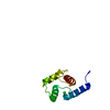
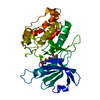
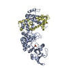
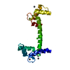
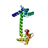
| Title | X-RAY STRUCTURE OF A DAP-KINASE CALMODULIN COMPLEX | ||||||
 Components Components |
| ||||||
 Keywords Keywords | TRANSFERASE/SIGNALING PROTEIN / TRANSFERASE-SIGNALING PROTEIN COMPLEX / TRANSFERASE SIGNALING PROTEIN COMPLEX / PHOSPHOPROTEIN / CALMODULIN-BINDING / DAPK / KINASE / ANK REPEAT / CALMODULIN / TRANSFERASE / ATP-BINDING | ||||||
| Function / homology |  Function and homology information Function and homology informationcellular response to hydroperoxide / regulation of response to tumor cell / positive regulation of autophagic cell death / DAPK1-calmodulin complex / : / establishment of protein localization to mitochondrial membrane / type 3 metabotropic glutamate receptor binding / defense response to tumor cell / Caspase activation via Dependence Receptors in the absence of ligand / regulation of NMDA receptor activity ...cellular response to hydroperoxide / regulation of response to tumor cell / positive regulation of autophagic cell death / DAPK1-calmodulin complex / : / establishment of protein localization to mitochondrial membrane / type 3 metabotropic glutamate receptor binding / defense response to tumor cell / Caspase activation via Dependence Receptors in the absence of ligand / regulation of NMDA receptor activity / CaM pathway / Cam-PDE 1 activation / Sodium/Calcium exchangers / syntaxin-1 binding / Calmodulin induced events / regulation of synaptic vesicle endocytosis / Reduction of cytosolic Ca++ levels / calcium/calmodulin-dependent protein kinase activity / CREB1 phosphorylation through the activation of CaMKII/CaMKK/CaMKIV cascasde / Activation of Ca-permeable Kainate Receptor / Loss of phosphorylation of MECP2 at T308 / CREB1 phosphorylation through the activation of Adenylate Cyclase / PKA activation / negative regulation of high voltage-gated calcium channel activity / CaMK IV-mediated phosphorylation of CREB / Glycogen breakdown (glycogenolysis) / positive regulation of cyclic-nucleotide phosphodiesterase activity / regulation of synaptic vesicle exocytosis / organelle localization by membrane tethering / negative regulation of calcium ion export across plasma membrane / CLEC7A (Dectin-1) induces NFAT activation / mitochondrion-endoplasmic reticulum membrane tethering / autophagosome membrane docking / regulation of cardiac muscle cell action potential / Activation of RAC1 downstream of NMDARs / response to corticosterone / positive regulation of DNA binding / positive regulation of ryanodine-sensitive calcium-release channel activity / nitric-oxide synthase binding / regulation of cell communication by electrical coupling involved in cardiac conduction / Negative regulation of NMDA receptor-mediated neuronal transmission / negative regulation of peptidyl-threonine phosphorylation / Synthesis of IP3 and IP4 in the cytosol / Unblocking of NMDA receptors, glutamate binding and activation / Phase 0 - rapid depolarisation / negative regulation of ryanodine-sensitive calcium-release channel activity / protein phosphatase activator activity / positive regulation of cysteine-type endopeptidase activity involved in apoptotic process / RHO GTPases activate PAKs / : / Ion transport by P-type ATPases / Long-term potentiation / Uptake and function of anthrax toxins / Regulation of MECP2 expression and activity / Calcineurin activates NFAT / adenylate cyclase binding / catalytic complex / DARPP-32 events / detection of calcium ion / extrinsic apoptotic signaling pathway via death domain receptors / regulation of cardiac muscle contraction / regulation of ryanodine-sensitive calcium-release channel activity / Smooth Muscle Contraction / cellular response to interferon-beta / RHO GTPases activate IQGAPs / regulation of cardiac muscle contraction by regulation of the release of sequestered calcium ion / calcium channel inhibitor activity / Protein methylation / phosphatidylinositol 3-kinase binding / eNOS activation / positive regulation of autophagy / enzyme regulator activity / activation of adenylate cyclase activity / regulation of release of sequestered calcium ion into cytosol by sarcoplasmic reticulum / Activation of AMPK downstream of NMDARs / Tetrahydrobiopterin (BH4) synthesis, recycling, salvage and regulation / : / Ion homeostasis / titin binding / regulation of calcium-mediated signaling / positive regulation of protein autophosphorylation / voltage-gated potassium channel complex / sperm midpiece / calcium channel complex / response to amphetamine / substantia nigra development / adenylate cyclase activator activity / Ras activation upon Ca2+ influx through NMDA receptor / nitric-oxide synthase regulator activity / regulation of heart rate / sarcomere / FCERI mediated Ca+2 mobilization / protein serine/threonine kinase activator activity / FCGR3A-mediated IL10 synthesis / VEGFR2 mediated vascular permeability / Antigen activates B Cell Receptor (BCR) leading to generation of second messengers / VEGFR2 mediated cell proliferation / regulation of cytokinesis / positive regulation of peptidyl-threonine phosphorylation / positive regulation of nitric-oxide synthase activity Similarity search - Function | ||||||
| Biological species |  HOMO SAPIENS (human) HOMO SAPIENS (human) | ||||||
| Method |  X-RAY DIFFRACTION / SYNCHROTRON / MOLECULAR REPLACEMENT / Resolution: 2.2 Å X-RAY DIFFRACTION / SYNCHROTRON / MOLECULAR REPLACEMENT / Resolution: 2.2 Å | ||||||
 Authors Authors | Kuper, J. / De Diego, I. / Lehmann, F. / Wilmanns, M. | ||||||
 Citation Citation | Journal: Sci.Signal / Year: 2010 Title: Molecular Basis of the Death-Associated Protein Kinase-Calcium/Calmodulin Regulator Complex. Authors: De Diego, I. / Kuper, J. / Bakalova, N. / Kursula, P. / Wilmanns, M. | ||||||
| History |
|
- Structure visualization
| Structure viewer | Molecule: MolmilJmol/JSmol |
|---|
- Downloads & links
-Download
| PDBx/mmCIF format | 2x0g.cif.gz | 111.7 KB | Display | PDBx/mmCIF format |
|---|---|---|---|---|
| PDB format | pdb2x0g.ent.gz | 84.7 KB | Display | PDB format |
| PDBx/mmJSON format | 2x0g.json.gz | Tree view | PDBx/mmJSON format | |
| Others |  Other downloads Other downloads |
-Validation report
| Summary document | 2x0g_validation.pdf.gz | 450.5 KB | Display | wwPDB validaton report |
|---|---|---|---|---|
| Full document | 2x0g_full_validation.pdf.gz | 457.3 KB | Display | |
| Data in XML | 2x0g_validation.xml.gz | 20 KB | Display | |
| Data in CIF | 2x0g_validation.cif.gz | 28.3 KB | Display | |
| Arichive directory | https://data.pdbj.org/pub/pdb/validation_reports/x0/2x0gftp://data.pdbj.org/pub/pdb/validation_reports/x0/2x0g | HTTPS FTP |
-Related structure data
| Related structure data |  1jksS S: Starting model for refinement |
|---|---|
| Similar structure data |


-Links
 PDBj
PDBj


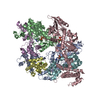
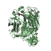
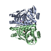
- Assembly
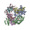
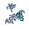
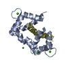
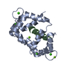
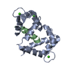
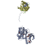
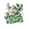
| Deposited unit | 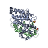
| ||||||||
|---|---|---|---|---|---|---|---|---|---|
| 1 |
| ||||||||
| Unit cell |
|
-Components
| #1: Protein | Mass: 38454.086 Da / Num. of mol.: 1 Fragment: CATALYTIC AND AUTOINHIBITORY DOMAIN, RESIDUES 1- 334 Source method: isolated from a genetically manipulated source Source: (gene. exp.) HOMO SAPIENS (human) / Plasmid: PETM11 / Production host:  References: UniProt: P53355, non-specific serine/threonine protein kinase | ||||
|---|---|---|---|---|---|
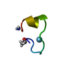
| #2: Protein | Mass: 16721.350 Da / Num. of mol.: 1 Source method: isolated from a genetically manipulated source Source: (gene. exp.) HOMO SAPIENS (human) / Plasmid: PETM8A / Production host: | ||||
| #3: Chemical |   Mass: 96.063 Da / Num. of mol.: 2 / Source method: obtained synthetically / Formula: SO4 Mass: 96.063 Da / Num. of mol.: 2 / Source method: obtained synthetically / Formula: SO4#4: Chemical | ChemComp-CA /   Mass: 40.078 Da / Num. of mol.: 4 / Source method: obtained synthetically / Formula: Ca Mass: 40.078 Da / Num. of mol.: 4 / Source method: obtained synthetically / Formula: Ca#5: Water | ChemComp-HOH / |  Mass: 18.015 Da / Num. of mol.: 177 / Source method: isolated from a natural source / Formula: H2O Mass: 18.015 Da / Num. of mol.: 177 / Source method: isolated from a natural source / Formula: H2O |
-Experimental details
-Experiment
| Experiment | Method: X-RAY DIFFRACTION / Number of used crystals: 1 |
|---|
- Sample preparation
| Crystal | Density Matthews: 2.43 Å3/Da / Density % sol: 41 % / Description: NONE |
|---|---|
| Crystal grow | pH: 7.2 Details: 0.17M AMMONIUM SULFATE, 25% (W/V) PEG 4000, 15% GLYCEROL, pH 7.2 |
-Data collection
| Diffraction | Mean temperature: 100 K |
|---|---|
| Diffraction source | Source: SYNCHROTRON / Site: ESRF  / Beamline: ID29 / Wavelength: 1.277 / Beamline: ID29 / Wavelength: 1.277 |
| Detector | Type: MARRESEARCH / Detector: CCD |
| Radiation | Protocol: SINGLE WAVELENGTH / Monochromatic (M) / Laue (L): M / Scattering type: x-ray |
| Radiation wavelength | Wavelength: 1.277 Å / Relative weight: 1 |
| Reflection | Resolution: 2.2→72.93 Å / Num. obs: 25927 / % possible obs: 100 % / Observed criterion σ(I): 0 / Redundancy: 3.4 % / Rmerge(I) obs: 0.09 / Net I/σ(I): 8.8 |
| Reflection shell | Resolution: 2.2→2.32 Å / Redundancy: 3.5 % / Rmerge(I) obs: 0.47 / Mean I/σ(I) obs: 2.6 / % possible all: 99.9 |
- Processing
| Software |
| ||||||||||||||||||||||||||||||||||||||||||||||||||||||||||||||||||||||||||||||||||||||||||||||||||||||||||||||||||||||||||||||||||||||||||||||||||||||||||||||||||||||||||||||||||||||
|---|---|---|---|---|---|---|---|---|---|---|---|---|---|---|---|---|---|---|---|---|---|---|---|---|---|---|---|---|---|---|---|---|---|---|---|---|---|---|---|---|---|---|---|---|---|---|---|---|---|---|---|---|---|---|---|---|---|---|---|---|---|---|---|---|---|---|---|---|---|---|---|---|---|---|---|---|---|---|---|---|---|---|---|---|---|---|---|---|---|---|---|---|---|---|---|---|---|---|---|---|---|---|---|---|---|---|---|---|---|---|---|---|---|---|---|---|---|---|---|---|---|---|---|---|---|---|---|---|---|---|---|---|---|---|---|---|---|---|---|---|---|---|---|---|---|---|---|---|---|---|---|---|---|---|---|---|---|---|---|---|---|---|---|---|---|---|---|---|---|---|---|---|---|---|---|---|---|---|---|---|---|---|---|
| Refinement | Method to determine structure: MOLECULAR REPLACEMENT Starting model: PDB ENTRY 1JKS Resolution: 2.2→52.41 Å / Cor.coef. Fo:Fc: 0.947 / Cor.coef. Fo:Fc free: 0.902 / SU B: 13.993 / SU ML: 0.173 / TLS residual ADP flag: LIKELY RESIDUAL / Cross valid method: THROUGHOUT / ESU R: 0.288 / ESU R Free: 0.236 / Stereochemistry target values: MAXIMUM LIKELIHOOD Details: HYDROGENS HAVE BEEN ADDED IN THE RIDING POSITIONS. U VALUES RESIDUAL ONLY.
| ||||||||||||||||||||||||||||||||||||||||||||||||||||||||||||||||||||||||||||||||||||||||||||||||||||||||||||||||||||||||||||||||||||||||||||||||||||||||||||||||||||||||||||||||||||||
| Solvent computation | Ion probe radii: 0.8 Å / Shrinkage radii: 0.8 Å / VDW probe radii: 1.2 Å / Solvent model: BABINET MODEL WITH MASK | ||||||||||||||||||||||||||||||||||||||||||||||||||||||||||||||||||||||||||||||||||||||||||||||||||||||||||||||||||||||||||||||||||||||||||||||||||||||||||||||||||||||||||||||||||||||
| Displacement parameters | Biso mean: 18.65 Å2
| ||||||||||||||||||||||||||||||||||||||||||||||||||||||||||||||||||||||||||||||||||||||||||||||||||||||||||||||||||||||||||||||||||||||||||||||||||||||||||||||||||||||||||||||||||||||
| Refinement step | Cycle: LAST / Resolution: 2.2→52.41 Å
| ||||||||||||||||||||||||||||||||||||||||||||||||||||||||||||||||||||||||||||||||||||||||||||||||||||||||||||||||||||||||||||||||||||||||||||||||||||||||||||||||||||||||||||||||||||||
| Refine LS restraints |
|