[English] 日本語
Yorodumi
- PDB-1sw8: Solution structure of the N-terminal domain of Human N60D calmodu... -

+
Open data


ID or keywords:

Loading...

-
Basic information

Entry
Database: PDB / ID: 1sw8
TitleSolution structure of the N-terminal domain of Human N60D calmodulin refined with paramagnetism based strategy
ComponentsCalmodulin
KeywordsCalcium-Binding Protein / calcium / calmodulin / EF-hand / lanthanide / Structural Proteomics in Europe / SPINE / Structural Genomics
Function / homology
Function and homology information


: / establishment of protein localization to mitochondrial membrane / type 3 metabotropic glutamate receptor binding / CaM pathway / Cam-PDE 1 activation / Sodium/Calcium exchangers / Calmodulin induced events / regulation of synaptic vesicle endocytosis / Reduction of cytosolic Ca++ levels / CREB1 phosphorylation through the activation of CaMKII/CaMKK/CaMKIV cascasde ...: / establishment of protein localization to mitochondrial membrane / type 3 metabotropic glutamate receptor binding / CaM pathway / Cam-PDE 1 activation / Sodium/Calcium exchangers / Calmodulin induced events / regulation of synaptic vesicle endocytosis / Reduction of cytosolic Ca++ levels / CREB1 phosphorylation through the activation of CaMKII/CaMKK/CaMKIV cascasde / Activation of Ca-permeable Kainate Receptor / Loss of phosphorylation of MECP2 at T308 / CREB1 phosphorylation through the activation of Adenylate Cyclase / PKA activation / negative regulation of high voltage-gated calcium channel activity / CaMK IV-mediated phosphorylation of CREB / Glycogen breakdown (glycogenolysis) / positive regulation of cyclic-nucleotide phosphodiesterase activity / regulation of synaptic vesicle exocytosis / organelle localization by membrane tethering / negative regulation of calcium ion export across plasma membrane / CLEC7A (Dectin-1) induces NFAT activation / mitochondrion-endoplasmic reticulum membrane tethering / autophagosome membrane docking / regulation of cardiac muscle cell action potential / Activation of RAC1 downstream of NMDARs / response to corticosterone / positive regulation of DNA binding / positive regulation of ryanodine-sensitive calcium-release channel activity / nitric-oxide synthase binding / regulation of cell communication by electrical coupling involved in cardiac conduction / Negative regulation of NMDA receptor-mediated neuronal transmission / negative regulation of peptidyl-threonine phosphorylation / Synthesis of IP3 and IP4 in the cytosol / Unblocking of NMDA receptors, glutamate binding and activation / Phase 0 - rapid depolarisation / negative regulation of ryanodine-sensitive calcium-release channel activity / protein phosphatase activator activity / RHO GTPases activate PAKs / : / Ion transport by P-type ATPases / Long-term potentiation / Uptake and function of anthrax toxins / Regulation of MECP2 expression and activity / Calcineurin activates NFAT / adenylate cyclase binding / catalytic complex / DARPP-32 events / detection of calcium ion / regulation of cardiac muscle contraction / regulation of ryanodine-sensitive calcium-release channel activity / Smooth Muscle Contraction / cellular response to interferon-beta / RHO GTPases activate IQGAPs / regulation of cardiac muscle contraction by regulation of the release of sequestered calcium ion / calcium channel inhibitor activity / Protein methylation / phosphatidylinositol 3-kinase binding / eNOS activation / enzyme regulator activity / activation of adenylate cyclase activity / regulation of release of sequestered calcium ion into cytosol by sarcoplasmic reticulum / Activation of AMPK downstream of NMDARs / Tetrahydrobiopterin (BH4) synthesis, recycling, salvage and regulation / : / Ion homeostasis / titin binding / regulation of calcium-mediated signaling / positive regulation of protein autophosphorylation / voltage-gated potassium channel complex / sperm midpiece / calcium channel complex / response to amphetamine / substantia nigra development / adenylate cyclase activator activity / Ras activation upon Ca2+ influx through NMDA receptor / nitric-oxide synthase regulator activity / regulation of heart rate / sarcomere / FCERI mediated Ca+2 mobilization / protein serine/threonine kinase activator activity / FCGR3A-mediated IL10 synthesis / VEGFR2 mediated vascular permeability / Antigen activates B Cell Receptor (BCR) leading to generation of second messengers / VEGFR2 mediated cell proliferation / regulation of cytokinesis / positive regulation of peptidyl-threonine phosphorylation / positive regulation of nitric-oxide synthase activity / Translocation of SLC2A4 (GLUT4) to the plasma membrane / spindle microtubule / mitochondrial membrane / RAF activation / positive regulation of receptor signaling pathway via JAK-STAT / Transcriptional activation of mitochondrial biogenesis / positive regulation of protein serine/threonine kinase activity / Stimuli-sensing channels / cellular response to type II interferon / spindle pole / synaptic vesicle membrane / response to calcium ion
Similarity search - Function
: / EF-hand / Recoverin; domain 1 / EF-hand domain pair / EF-hand, calcium binding motif / EF-Hand 1, calcium-binding site / EF-hand calcium-binding domain. / EF-hand calcium-binding domain profile. / EF-hand domain / EF-hand domain pair ...: / EF-hand / Recoverin; domain 1 / EF-hand domain pair / EF-hand, calcium binding motif / EF-Hand 1, calcium-binding site / EF-hand calcium-binding domain. / EF-hand calcium-binding domain profile. / EF-hand domain / EF-hand domain pair / Orthogonal Bundle / Mainly Alpha
Similarity search - Domain/homology
Calmodulin-1 / Calmodulin-3
Similarity search - Component
Biological speciesHomo sapiens (human)
MethodSOLUTION NMR / distance geometry, simulated annealing, torsion angle dynamics
AuthorsBertini, I. / Del Bianco, C. / Gelis, I. / Katsaros, N. / Luchinat, C. / Parigi, G. / Peana, M. / Provenzani, A. / Zoroddu, M.A. / Structural Proteomics in Europe (SPINE)
CitationJournal: Proc.Natl.Acad.Sci.USA / Year: 2004
Title: Experimentally exploring the conformational space sampled by domain reorientation in calmodulin
Authors: Bertini, I. / Del Bianco, C. / Gelis, I. / Katsaros, N. / Luchinat, C. / Parigi, G. / Peana, M. / Provenzani, A. / Zoroddu, M.A.
History
DepositionMar 30, 2004Deposition site: RCSB / Processing site: RCSB
Revision 1.0Apr 6, 2004Provider: repository / Type: Initial release
Revision 1.1Apr 30, 2008Group: Version format compliance
Revision 1.2Jul 13, 2011Group: Version format compliance
Revision 1.3Oct 27, 2021Group: Data collection / Database references / Derived calculations
Category: database_2 / pdbx_nmr_spectrometer ...database_2 / pdbx_nmr_spectrometer / pdbx_struct_assembly / pdbx_struct_conn_angle / pdbx_struct_oper_list / struct_conn / struct_ref_seq_dif / struct_site
Item: _database_2.pdbx_DOI / _database_2.pdbx_database_accession ..._database_2.pdbx_DOI / _database_2.pdbx_database_accession / _pdbx_nmr_spectrometer.model / _pdbx_struct_conn_angle.ptnr1_auth_comp_id / _pdbx_struct_conn_angle.ptnr1_auth_seq_id / _pdbx_struct_conn_angle.ptnr1_label_atom_id / _pdbx_struct_conn_angle.ptnr1_label_comp_id / _pdbx_struct_conn_angle.ptnr1_label_seq_id / _pdbx_struct_conn_angle.ptnr3_auth_comp_id / _pdbx_struct_conn_angle.ptnr3_auth_seq_id / _pdbx_struct_conn_angle.ptnr3_label_atom_id / _pdbx_struct_conn_angle.ptnr3_label_comp_id / _pdbx_struct_conn_angle.ptnr3_label_seq_id / _pdbx_struct_conn_angle.value / _struct_conn.pdbx_dist_value / _struct_conn.ptnr1_auth_comp_id / _struct_conn.ptnr1_auth_seq_id / _struct_conn.ptnr1_label_asym_id / _struct_conn.ptnr1_label_atom_id / _struct_conn.ptnr1_label_comp_id / _struct_conn.ptnr1_label_seq_id / _struct_conn.ptnr2_auth_comp_id / _struct_conn.ptnr2_auth_seq_id / _struct_conn.ptnr2_label_asym_id / _struct_conn.ptnr2_label_atom_id / _struct_conn.ptnr2_label_comp_id / _struct_conn.ptnr2_label_seq_id / _struct_ref_seq_dif.details / _struct_site.pdbx_auth_asym_id / _struct_site.pdbx_auth_comp_id / _struct_site.pdbx_auth_seq_id
Revision 1.4May 22, 2024Group: Data collection / Category: chem_comp_atom / chem_comp_bond

-
Structure visualization

Structure viewerMolecule:
MolmilJmol/JSmol

Downloads & links

-
Assembly

Deposited unit
A: Calmodulin
hetero molecules


Theoretical massNumber of molelcules
Total (without water)8,8813
Polymers8,8011
Non-polymers802
Water00
1


  • Idetical with deposited unit
  • defined by author
TypeNameSymmetry operationNumber
identity operation1_5551
NMR ensembles
DataCriteria
Number of conformers (submitted / calculated)20 / 200structures with the lowest energy
RepresentativeModel #1lowest energy

-
Components

#1: Protein Calmodulin


Mass: 8800.708 Da / Num. of mol.: 1 / Fragment: N-terminal domain / Mutation: N60D
Source method: isolated from a genetically manipulated source
Source: (gene. exp.) Homo sapiens (human) / Production host: Escherichia coli (E. coli) / References: UniProt: P62158, UniProt: P0DP23*PLUS
#2: Chemical ChemComp-CA / CALCIUM ION


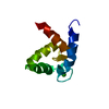
Mass: 40.078 Da / Num. of mol.: 2 / Source method: obtained synthetically / Formula: Ca

-
Experimental details

-
Experiment

ExperimentMethod: SOLUTION NMR
NMR experiment
Conditions-IDExperiment-IDSolution-IDType
1123D 13C-separated NOESY
1213D 15N-separated NOESY
1312D NOESY
NMR detailsText: The structure was calculated using dimagnetic, dihedral angle restraints and pseudocontact shifts

-
Sample preparation

Details
Solution-IDContentsSolvent system
11 mM Calmodulin U-15N, 20 mM MES, 400 mM KCl, 90% H20, 10% D2090%H20, 10% D20
21 mM Calmodulin U-15N-13C, 20 mM MES, 400 mM KCl, 90% H20, 10% D2090%H20, 10% D20
Sample conditionsIonic strength: 400 mM KCl / pH: 6.5 / Pressure: ambient / Temperature: 300 K

-
NMR measurement

RadiationProtocol: SINGLE WAVELENGTH / Monochromatic (M) / Laue (L): M
Radiation wavelengthRelative weight: 1
NMR spectrometer
TypeManufacturerModelField strength (MHz)Spectrometer-ID
Bruker AVANCEBrukerAVANCE7001
Bruker AVANCEBrukerAVANCE8002

-
Processing

NMR software
NameVersionClassification
XWINNMR 3.13.1collection
XWINNMR 3.13.1processing
XEASY1.3data analysis
DYANAstructure solution
DYANArefinement
RefinementMethod: distance geometry, simulated annealing, torsion angle dynamics
Software ordinal: 1 / Details: refinement with pseudocontact shifts
NMR representativeSelection criteria: lowest energy
NMR ensembleConformer selection criteria: structures with the lowest energy
Conformers calculated total number: 200 / Conformers submitted total number: 20

+
About Yorodumi

-
News

-
Feb 9, 2022. New format data for meta-information of EMDB entries

New format data for meta-information of EMDB entries

  • Version 3 of the EMDB header file is now the official format.
  • The previous official version 1.9 will be removed from the archive.

Related info.:EMDB header

External links:wwPDB to switch to version 3 of the EMDB data model

-
Aug 12, 2020. Covid-19 info

Covid-19 info

URL: https://pdbjlc1.pdbj.org/emnavi/covid19.php

New page: Covid-19 featured information page in EM Navigator.

Related info.:Covid-19 info / Mar 5, 2020. Novel coronavirus structure data

+
Mar 5, 2020. Novel coronavirus structure data

Novel coronavirus structure data

Related info.:Yorodumi Speices / Aug 12, 2020. Covid-19 info

External links:COVID-19 featured content - PDBj / Molecule of the Month (242):Coronavirus Proteases

+
Jan 31, 2019. EMDB accession codes are about to change! (news from PDBe EMDB page)

EMDB accession codes are about to change! (news from PDBe EMDB page)

  • The allocation of 4 digits for EMDB accession codes will soon come to an end. Whilst these codes will remain in use, new EMDB accession codes will include an additional digit and will expand incrementally as the available range of codes is exhausted. The current 4-digit format prefixed with “EMD-” (i.e. EMD-XXXX) will advance to a 5-digit format (i.e. EMD-XXXXX), and so on. It is currently estimated that the 4-digit codes will be depleted around Spring 2019, at which point the 5-digit format will come into force.
  • The EM Navigator/Yorodumi systems omit the EMD- prefix.

Related info.:Q: What is EMD? / ID/Accession-code notation in Yorodumi/EM Navigator

External links:EMDB Accession Codes are Changing Soon! / Contact to PDBj

+
Jul 12, 2017. Major update of PDB

Major update of PDB

  • wwPDB released updated PDB data conforming to the new PDBx/mmCIF dictionary.
  • This is a major update changing the version number from 4 to 5, and with Remediation, in which all the entries are updated.
  • In this update, many items about electron microscopy experimental information are reorganized (e.g. em_software).
  • Now, EM Navigator and Yorodumi are based on the updated data.

External links:wwPDB Remediation / Enriched Model Files Conforming to OneDep Data Standards Now Available in the PDB FTP Archive

-
Yorodumi

Thousand views of thousand structures

  • Yorodumi is a browser for structure data from EMDB, PDB, SASBDB, etc.
  • This page is also the successor to EM Navigator detail page, and also detail information page/front-end page for Omokage search.
  • The word "yorodu" (or yorozu) is an old Japanese word meaning "ten thousand". "mi" (miru) is to see.

Related info.:EMDB / PDB / SASBDB / Comparison of 3 databanks / Yorodumi Search / Aug 31, 2016. New EM Navigator & Yorodumi / Yorodumi Papers / Jmol/JSmol / Function and homology information / Changes in new EM Navigator and Yorodumi

Read more