 Movie
Movie Controller

[English] 日本語
 Yorodumi
- PDB-1ayc: CRYSTAL STRUCTURES OF PEPTIDE COMPLEXES OF THE AMINO-TERMINAL SH2... -
+ Open data

- Basic information
| Entry | Database: PDB / ID: 1ayc | ||||||
|---|---|---|---|---|---|---|---|
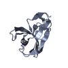
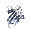
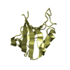
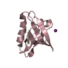
| Title | CRYSTAL STRUCTURES OF PEPTIDE COMPLEXES OF THE AMINO-TERMINAL SH2 DOMAIN OF THE SYP TYROSINE PHOSPHATASE | ||||||
 Components Components |
| ||||||
 Keywords Keywords | HYDROLASE(SH2 DOMAIN) | ||||||
| Function / homology |  Function and homology information Function and homology informationmetanephric mesenchymal cell migration / metanephric glomerular mesangium development / response to ceramide / Signaling by PDGF / PI-3K cascade:FGFR3 / PI-3K cascade:FGFR4 / Co-inhibition by BTLA / PI-3K cascade:FGFR1 / PI-3K cascade:FGFR2 / platelet-derived growth factor receptor activity ...metanephric mesenchymal cell migration / metanephric glomerular mesangium development / response to ceramide / Signaling by PDGF / PI-3K cascade:FGFR3 / PI-3K cascade:FGFR4 / Co-inhibition by BTLA / PI-3K cascade:FGFR1 / PI-3K cascade:FGFR2 / platelet-derived growth factor receptor activity / cell migration involved in coronary angiogenesis / metanephric glomerular mesangial cell proliferation involved in metanephros development / FRS-mediated FGFR3 signaling / FRS-mediated FGFR4 signaling / MET activates PTPN11 / Regulation of IFNA/IFNB signaling / FRS-mediated FGFR1 signaling / FRS-mediated FGFR2 signaling / platelet-derived growth factor beta-receptor activity / cell migration involved in vasculogenesis / Tie2 Signaling / positive regulation of metanephric mesenchymal cell migration by platelet-derived growth factor receptor-beta signaling pathway / PECAM1 interactions / Interleukin-20 family signaling / Signaling by CSF3 (G-CSF) / cardiac vascular smooth muscle cell differentiation / GAB1 signalosome / PI3K Cascade / MAPK3 (ERK1) activation / MAPK1 (ERK2) activation / Co-inhibition by CTLA4 / Regulation of RUNX1 Expression and Activity / lung growth / metanephric glomerular capillary formation / smooth muscle cell chemotaxis / Interleukin-6 signaling / Downstream signal transduction / GPVI-mediated activation cascade / Co-inhibition by PD-1 / Signaling by SCF-KIT / Negative regulation of FGFR3 signaling / Negative regulation of FGFR4 signaling / Negative regulation of FGFR1 signaling / Negative regulation of FGFR2 signaling / Spry regulation of FGF signaling / glycosaminoglycan biosynthetic process / Activation of IRF3, IRF7 mediated by TBK1, IKKε (IKBKE) / aorta morphogenesis / platelet-derived growth factor binding / positive regulation of cell proliferation by VEGF-activated platelet derived growth factor receptor signaling pathway / negative regulation of hormone secretion / positive regulation of hepatic stellate cell activation / positive regulation of signal transduction / PIP3 activates AKT signaling / vascular endothelial growth factor binding / negative regulation of cortisol secretion / intestinal epithelial cell migration / microvillus organization / negative regulation of growth hormone secretion / RAF/MAP kinase cascade / Platelet sensitization by LDL / retina vasculature development in camera-type eye / atrioventricular canal development / ruffle assembly / PI5P, PP2A and IER3 Regulate PI3K/AKT Signaling / genitalia development / Interleukin-3, Interleukin-5 and GM-CSF signaling / smooth muscle tissue development / RET signaling / cardiac myofibril assembly / tissue homeostasis / negative regulation of neutrophil activation / cerebellar cortex formation / positive regulation of chemotaxis / phospholipase C activator activity / positive regulation of hormone secretion / regulation of protein export from nucleus / positive regulation of lipopolysaccharide-mediated signaling pathway / negative regulation of cell adhesion mediated by integrin / positive regulation of ossification / platelet-derived growth factor receptor binding / regulation of cell adhesion mediated by integrin / hormone metabolic process / negative regulation of chondrocyte differentiation / face morphogenesis / platelet formation / skeletal system morphogenesis / organ growth / ERBB signaling pathway / regulation of peptidyl-tyrosine phosphorylation / triglyceride metabolic process / adrenal gland development / megakaryocyte development / positive regulation of smooth muscle cell migration / positive regulation of DNA biosynthetic process / negative regulation of T cell activation / peptide hormone receptor binding / platelet-derived growth factor receptor-beta signaling pathway / negative regulation of type I interferon production / blood vessel development Similarity search - Function | ||||||
| Biological species |  | ||||||
| Method |  X-RAY DIFFRACTION / Resolution: 2.3 Å X-RAY DIFFRACTION / Resolution: 2.3 Å | ||||||
 Authors Authors | Lee, C.-H. / Kuriyan, J. | ||||||
 Citation Citation | Journal: Structure / Year: 1994 Title: Crystal structures of peptide complexes of the amino-terminal SH2 domain of the Syp tyrosine phosphatase. Authors: Lee, C.H. / Kominos, D. / Jacques, S. / Margolis, B. / Schlessinger, J. / Shoelson, S.E. / Kuriyan, J. #1: Journal: Cell(Cambridge,Mass.) / Year: 1993Title: Binding of a High Affinity Phosphotyrosyl Peptide to the Src Sh2 Domain: Crystal Structures of the Complexed and Peptide-Free Forms Authors: Waksman, G. / Shoelson, S.E. / Pant, N. / Cowburn, D. / Kuriyan, J. #2: Journal: Curr.Opin.Struct.Biol. / Year: 1993Title: Structures of Sh2 and SH3 Domains Authors: Kuriyan, J. / Cowburn, D. #3: Journal: Nature / Year: 1992Title: Crystal Structure of the Phosphotyrosine Recognition Domain Sh2 of V-Src Complexed with Tyrosine-Phosphorylated Peptides Authors: Waksman, G. / Kominos, D. / Robertson, S.R. / Pant, N. / Baltimore, D. / Birge, R.B. / Cowburn, D. / Hanafusa, H. / Mayer, B.J. / Overduin, M. / Resh, M.D. / Rios, C.B. / Silverman, L. / Kuriyan, J. | ||||||
| History |
|
- Structure visualization
| Structure viewer | Molecule: MolmilJmol/JSmol |
|---|
- Downloads & links
-Download
| PDBx/mmCIF format | 1ayc.cif.gz | 34.8 KB | Display | PDBx/mmCIF format |
|---|---|---|---|---|
| PDB format | pdb1ayc.ent.gz | 23.4 KB | Display | PDB format |
| PDBx/mmJSON format | 1ayc.json.gz | Tree view | PDBx/mmJSON format | |
| Others |  Other downloads Other downloads |
-Validation report
| Arichive directory | https://data.pdbj.org/pub/pdb/validation_reports/ay/1aycftp://data.pdbj.org/pub/pdb/validation_reports/ay/1ayc | HTTPS FTP |
|---|
-Related structure data


-Links
 PDBj
PDBj


- Assembly
| Deposited unit | 
| ||||||||
|---|---|---|---|---|---|---|---|---|---|
| 1 |
| ||||||||
| Unit cell |
|
-Components
| #1: Protein | Mass: 11528.979 Da / Num. of mol.: 1 Source method: isolated from a genetically manipulated source Source: (gene. exp.) |
|---|---|
| #2: Protein/peptide | Mass: 1228.226 Da / Num. of mol.: 1 Source method: isolated from a genetically manipulated source Source: (gene. exp.) |
| #3: Water | ChemComp-HOH /  Mass: 18.015 Da / Num. of mol.: 63 / Source method: isolated from a natural source / Formula: H2O Mass: 18.015 Da / Num. of mol.: 63 / Source method: isolated from a natural source / Formula: H2O |
| Has protein modification | Y |
-Experimental details
-Experiment
| Experiment | Method: X-RAY DIFFRACTION |
|---|
- Sample preparation
| Crystal | Density Matthews: 2.83 Å3/Da / Density % sol: 56.55 % | |||||||||||||||||||||||||
|---|---|---|---|---|---|---|---|---|---|---|---|---|---|---|---|---|---|---|---|---|---|---|---|---|---|---|
| Crystal grow | *PLUS pH: 6 / Method: vapor diffusion, hanging drop | |||||||||||||||||||||||||
| Components of the solutions | *PLUS
|
-Data collection
| Radiation | Scattering type: x-ray |
|---|---|
| Radiation wavelength | Relative weight: 1 |
| Reflection | *PLUS Highest resolution: 2.3 Å / Lowest resolution: 30 Å / Num. obs: 6914 / % possible obs: 96 % / Num. measured all: 43918 / Rmerge(I) obs: 0.054 |
| Reflection shell | *PLUS Highest resolution: 2.3 Å / Lowest resolution: 2.35 Å / % possible obs: 90.9 % |
- Processing
| Software |
| ||||||||||||||||||||||||||||||||||||||||||||||||||||||||||||
|---|---|---|---|---|---|---|---|---|---|---|---|---|---|---|---|---|---|---|---|---|---|---|---|---|---|---|---|---|---|---|---|---|---|---|---|---|---|---|---|---|---|---|---|---|---|---|---|---|---|---|---|---|---|---|---|---|---|---|---|---|---|
| Refinement | Resolution: 2.3→6 Å / σ(F): 2 /
| ||||||||||||||||||||||||||||||||||||||||||||||||||||||||||||
| Refinement step | Cycle: LAST / Resolution: 2.3→6 Å
| ||||||||||||||||||||||||||||||||||||||||||||||||||||||||||||
| Refine LS restraints |
| ||||||||||||||||||||||||||||||||||||||||||||||||||||||||||||
| Software | *PLUS Name: X-PLOR / Classification: refinement | ||||||||||||||||||||||||||||||||||||||||||||||||||||||||||||
| Refinement | *PLUS Rfactor obs: 0.189 | ||||||||||||||||||||||||||||||||||||||||||||||||||||||||||||
| Solvent computation | *PLUS | ||||||||||||||||||||||||||||||||||||||||||||||||||||||||||||
| Displacement parameters | *PLUS | ||||||||||||||||||||||||||||||||||||||||||||||||||||||||||||
| Refine LS restraints | *PLUS Type: x_angle_d / Dev ideal: 2.9 |
