 Movie
Movie Controller

[English] 日本語
 Yorodumi
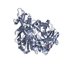
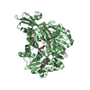
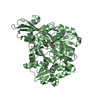
- PDB-6m2t: The crystal structure of benzoate coenzyme A ligase double mutant... -
+ Open data

- Basic information
| Entry | Database: PDB / ID: 6m2t | |||||||||
|---|---|---|---|---|---|---|---|---|---|---|
| Title | The crystal structure of benzoate coenzyme A ligase double mutant (H333A/I334A) in complex with 2-methyl-thiazole-5 carboxylate-AMP | |||||||||
 Components Components | Benzoate-coenzyme A ligase | |||||||||
 Keywords Keywords | LIGASE | |||||||||
| Function / homology |  Function and homology information Function and homology informationacid-thiol ligase activity / CoA-ligase activity / secondary metabolite biosynthetic process / ATP binding Similarity search - Function | |||||||||
| Biological species |  Rhodopseudomonas palustris (phototrophic) Rhodopseudomonas palustris (phototrophic) | |||||||||
| Method |  X-RAY DIFFRACTION / SYNCHROTRON / MOLECULAR REPLACEMENT / Resolution: 2.14 Å X-RAY DIFFRACTION / SYNCHROTRON / MOLECULAR REPLACEMENT / Resolution: 2.14 Å | |||||||||
 Authors Authors | Li, T.L. / Adhikari, K. | |||||||||
 Citation Citation | Journal: To Be Published Title: The crystal structure of benzoate coenzyme A ligase double mutant (H333A/I334A) in complex with 2-methyl-thiazole-5 carboxylate-AMP Authors: Li, T.L. / Adhikari, K. / Malek, Z.S. | |||||||||
| History |
|
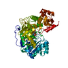
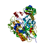
- Structure visualization
| Structure viewer | Molecule: MolmilJmol/JSmol |
|---|
- Downloads & links
-Download
| PDBx/mmCIF format | 6m2t.cif.gz | 425.4 KB | Display | PDBx/mmCIF format |
|---|---|---|---|---|
| PDB format | pdb6m2t.ent.gz | 341.9 KB | Display | PDB format |
| PDBx/mmJSON format | 6m2t.json.gz | Tree view | PDBx/mmJSON format | |
| Others |  Other downloads Other downloads |
-Validation report
| Arichive directory | https://data.pdbj.org/pub/pdb/validation_reports/m2/6m2tftp://data.pdbj.org/pub/pdb/validation_reports/m2/6m2t | HTTPS FTP |
|---|
-Related structure data
| Related structure data | 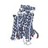 6m20S S: Starting model for refinement |
|---|---|
| Similar structure data |


-Links
 PDBj
PDBj


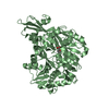
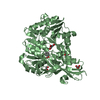
- Assembly
| Deposited unit | 
| ||||||||
|---|---|---|---|---|---|---|---|---|---|
| 1 | 
| ||||||||
| 2 | 
| ||||||||
| 3 | 
| ||||||||
| 4 | 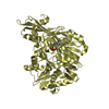
| ||||||||
| Unit cell |
|
-Components
| #1: Protein | Mass: 56683.098 Da / Num. of mol.: 4 / Mutation: T83A, H333A, I334A, G341D Source method: isolated from a genetically manipulated source Source: (gene. exp.) Rhodopseudomonas palustris (phototrophic)Gene: badA / Production host: Rhodopseudomonas palustris (phototrophic) / References: UniProt: Q93TK0#2: Chemical | ChemComp-AMP /   Mass: 347.221 Da / Num. of mol.: 4 / Source method: obtained synthetically / Formula: C10H14N5O7P / Feature type: SUBJECT OF INVESTIGATION / Comment: AMP*YM Mass: 347.221 Da / Num. of mol.: 4 / Source method: obtained synthetically / Formula: C10H14N5O7P / Feature type: SUBJECT OF INVESTIGATION / Comment: AMP*YM#3: Chemical | ChemComp-6V9 /   Type: peptide linking / Mass: 143.164 Da / Num. of mol.: 4 / Source method: obtained synthetically / Formula: C5H5NO2S / Feature type: SUBJECT OF INVESTIGATION Type: peptide linking / Mass: 143.164 Da / Num. of mol.: 4 / Source method: obtained synthetically / Formula: C5H5NO2S / Feature type: SUBJECT OF INVESTIGATION#4: Water | ChemComp-HOH / |  Mass: 18.015 Da / Num. of mol.: 1377 / Source method: isolated from a natural source / Formula: H2O Mass: 18.015 Da / Num. of mol.: 1377 / Source method: isolated from a natural source / Formula: H2OHas ligand of interest | Y | |
|---|
-Experimental details
-Experiment
| Experiment | Method: X-RAY DIFFRACTION / Number of used crystals: 1 |
|---|
- Sample preparation
| Crystal | Density Matthews: 2.32 Å3/Da / Density % sol: 47.05 % |
|---|---|
| Crystal grow | Temperature: 293 K / Method: vapor diffusion, hanging drop / pH: 6.5 / Details: PEG 4000, 0.1 M sodium chloride, 0.2 M MES |
-Data collection
| Diffraction | Mean temperature: 200 K / Serial crystal experiment: N |
|---|---|
| Diffraction source | Source: SYNCHROTRON / Site: NSRRC  / Beamline: BL15A1 / Wavelength: 1 Å / Beamline: BL15A1 / Wavelength: 1 Å |
| Detector | Type: RAYONIX MX300HE / Detector: CCD / Date: Jun 19, 2019 |
| Radiation | Protocol: SINGLE WAVELENGTH / Monochromatic (M) / Laue (L): M / Scattering type: x-ray |
| Radiation wavelength | Wavelength: 1 Å / Relative weight: 1 |
| Reflection | Resolution: 2.138→27.783 Å / Num. obs: 113090 / % possible obs: 99.4 % / Redundancy: 3.8 % / Rmerge(I) obs: 0.2 / Net I/σ(I): 25.33 |
| Reflection shell | Resolution: 2.14→2.22 Å / Rmerge(I) obs: 0.2 / Num. unique obs: 113090 / % possible all: 100 |
- Processing
| Software |
| |||||||||||||||||||||||||||||||||||||||||||||||||||||||||||||||||||||||||||||||||||||||||||||||||||||||||
|---|---|---|---|---|---|---|---|---|---|---|---|---|---|---|---|---|---|---|---|---|---|---|---|---|---|---|---|---|---|---|---|---|---|---|---|---|---|---|---|---|---|---|---|---|---|---|---|---|---|---|---|---|---|---|---|---|---|---|---|---|---|---|---|---|---|---|---|---|---|---|---|---|---|---|---|---|---|---|---|---|---|---|---|---|---|---|---|---|---|---|---|---|---|---|---|---|---|---|---|---|---|---|---|---|---|---|
| Refinement | Method to determine structure: MOLECULAR REPLACEMENT Starting model: 6M20 Resolution: 2.14→27.78 Å / SU ML: 0.19 / Cross valid method: NONE / σ(F): 1.34 / Phase error: 20.42 / Stereochemistry target values: ML
| |||||||||||||||||||||||||||||||||||||||||||||||||||||||||||||||||||||||||||||||||||||||||||||||||||||||||
| Solvent computation | Shrinkage radii: 0.9 Å / VDW probe radii: 1.11 Å / Solvent model: FLAT BULK SOLVENT MODEL | |||||||||||||||||||||||||||||||||||||||||||||||||||||||||||||||||||||||||||||||||||||||||||||||||||||||||
| Refinement step | Cycle: LAST / Resolution: 2.14→27.78 Å
| |||||||||||||||||||||||||||||||||||||||||||||||||||||||||||||||||||||||||||||||||||||||||||||||||||||||||
| Refine LS restraints |
| |||||||||||||||||||||||||||||||||||||||||||||||||||||||||||||||||||||||||||||||||||||||||||||||||||||||||
| LS refinement shell |
|
