 Movie
Movie Controller

[English] 日本語
 Yorodumi
- PDB-6m20: Crystal structure of Plasmodium falciparum hexose transporter PfH... -
+ Open data

- Basic information
| Entry | Database: PDB / ID: 6m20 | |||||||||
|---|---|---|---|---|---|---|---|---|---|---|
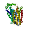
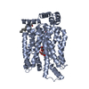
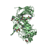
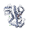
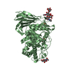
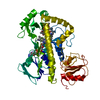
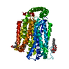
| Title | Crystal structure of Plasmodium falciparum hexose transporter PfHT1 bound with glucose | |||||||||
 Components Components | Hexose transporter 1 | |||||||||
 Keywords Keywords | TRANSPORT PROTEIN / MFS / hexose transporter / occluded state / Plasmodium falciparum | |||||||||
| Function / homology |  Function and homology information Function and homology information | |||||||||
| Biological species |  | |||||||||
| Method |  X-RAY DIFFRACTION / SYNCHROTRON / MOLECULAR REPLACEMENT / Resolution: 2.6 Å X-RAY DIFFRACTION / SYNCHROTRON / MOLECULAR REPLACEMENT / Resolution: 2.6 Å | |||||||||
 Authors Authors | Jiang, X. / Yuan, Y.Y. / Zhang, S. / Wang, N. / Yan, C.Y. / Yan, N. | |||||||||
| Funding support |  China, 2items China, 2items
| |||||||||
 Citation Citation | Journal: Cell / Year: 2020 Title: Structural Basis for Blocking Sugar Uptake into the Malaria Parasite Plasmodium falciparum. Authors: Jiang, X. / Yuan, Y. / Huang, J. / Zhang, S. / Luo, S. / Wang, N. / Pu, D. / Zhao, N. / Tang, Q. / Hirata, K. / Yang, X. / Jiao, Y. / Sakata-Kato, T. / Wu, J.W. / Yan, C. / Kato, N. / Yin, H. / Yan, N. | |||||||||
| History |
|
- Structure visualization
| Structure viewer | Molecule: MolmilJmol/JSmol |
|---|
- Downloads & links
-Download
| PDBx/mmCIF format | 6m20.cif.gz | 379.7 KB | Display | PDBx/mmCIF format |
|---|---|---|---|---|
| PDB format | pdb6m20.ent.gz | 310.8 KB | Display | PDB format |
| PDBx/mmJSON format | 6m20.json.gz | Tree view | PDBx/mmJSON format | |
| Others |  Other downloads Other downloads |
-Validation report
| Arichive directory | https://data.pdbj.org/pub/pdb/validation_reports/m2/6m20ftp://data.pdbj.org/pub/pdb/validation_reports/m2/6m20 | HTTPS FTP |
|---|
-Related structure data
| Related structure data | 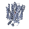 6m2lC 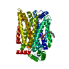 4zw9S S: Starting model for refinement C: citing same article ( |
|---|---|
| Similar structure data |
-Links
 PDBj
PDBj


- Assembly
| Deposited unit | 
| ||||||||
|---|---|---|---|---|---|---|---|---|---|
| 1 | 
| ||||||||
| 2 | 
| ||||||||
| 3 | 
| ||||||||
| 4 | 
| ||||||||
| Unit cell |
|
-Components
| #1: Protein | Mass: 56468.301 Da / Num. of mol.: 4 Source method: isolated from a genetically manipulated source Source: (gene. exp.) Gene: ht1 / Production host:   Spodoptera frugiperda (fall armyworm) / References: UniProt: O97467 Spodoptera frugiperda (fall armyworm) / References: UniProt: O97467#2: Sugar | ChemComp-BGC /   Type: D-saccharide, beta linking / Mass: 180.156 Da / Num. of mol.: 4 / Source method: isolated from a natural source / Formula: C6H12O6 / Feature type: SUBJECT OF INVESTIGATION Type: D-saccharide, beta linking / Mass: 180.156 Da / Num. of mol.: 4 / Source method: isolated from a natural source / Formula: C6H12O6 / Feature type: SUBJECT OF INVESTIGATION#3: Sugar | ChemComp-BNG /   Type: D-saccharide / Mass: 306.395 Da / Num. of mol.: 13 / Source method: isolated from a natural source / Formula: C15H30O6 / Comment: detergent*YM Type: D-saccharide / Mass: 306.395 Da / Num. of mol.: 13 / Source method: isolated from a natural source / Formula: C15H30O6 / Comment: detergent*YM#4: Water | ChemComp-HOH / |  Mass: 18.015 Da / Num. of mol.: 46 / Source method: isolated from a natural source / Formula: H2O Mass: 18.015 Da / Num. of mol.: 46 / Source method: isolated from a natural source / Formula: H2OHas ligand of interest | Y | Has protein modification | Y | |
|---|
-Experimental details
-Experiment
| Experiment | Method: X-RAY DIFFRACTION / Number of used crystals: 1 |
|---|
- Sample preparation
| Crystal | Density Matthews: 3.26 Å3/Da / Density % sol: 62.28 % |
|---|---|
| Crystal grow | Temperature: 291 K / Method: vapor diffusion, hanging drop / pH: 5.7 Details: 0.1 M NaCl, 0.1 M MES pH 5.7, 26% (w/v) PEG 400 and 2% (w/v) Benzamidine hydrochloride |
-Data collection
| Diffraction | Mean temperature: 80 K / Serial crystal experiment: N |
|---|---|
| Diffraction source | Source: SYNCHROTRON / Site: SPring-8  / Beamline: BL32XU / Wavelength: 1 Å / Beamline: BL32XU / Wavelength: 1 Å |
| Detector | Type: DECTRIS EIGER X 9M / Detector: PIXEL / Date: Nov 29, 2017 |
| Radiation | Protocol: SINGLE WAVELENGTH / Monochromatic (M) / Laue (L): M / Scattering type: x-ray |
| Radiation wavelength | Wavelength: 1 Å / Relative weight: 1 |
| Reflection | Resolution: 2.6→50 Å / Num. obs: 84480 / % possible obs: 98.8 % / Redundancy: 3.43 % / CC1/2: 0.998 / Net I/σ(I): 7.89 |
| Reflection shell | Resolution: 2.6→2.7 Å / Num. unique obs: 8948 / CC1/2: 0.666 |
- Processing
| Software |
| ||||||||||||||||||||||||||||||||||||||||||||||||||||||||||||||||||||||||||||||||||||||||||||||||||||||||||||||||||||||||||||||||||||||||||||||||||||||||||||||||||||||||||||||||||||||||||
|---|---|---|---|---|---|---|---|---|---|---|---|---|---|---|---|---|---|---|---|---|---|---|---|---|---|---|---|---|---|---|---|---|---|---|---|---|---|---|---|---|---|---|---|---|---|---|---|---|---|---|---|---|---|---|---|---|---|---|---|---|---|---|---|---|---|---|---|---|---|---|---|---|---|---|---|---|---|---|---|---|---|---|---|---|---|---|---|---|---|---|---|---|---|---|---|---|---|---|---|---|---|---|---|---|---|---|---|---|---|---|---|---|---|---|---|---|---|---|---|---|---|---|---|---|---|---|---|---|---|---|---|---|---|---|---|---|---|---|---|---|---|---|---|---|---|---|---|---|---|---|---|---|---|---|---|---|---|---|---|---|---|---|---|---|---|---|---|---|---|---|---|---|---|---|---|---|---|---|---|---|---|---|---|---|---|---|---|
| Refinement | Method to determine structure: MOLECULAR REPLACEMENT Starting model: 4ZW9 Resolution: 2.6→20 Å / FOM work R set: 0.7162 / SU ML: 0.38 / Cross valid method: THROUGHOUT / σ(F): 1.35 / Phase error: 33.96 / Stereochemistry target values: ML
| ||||||||||||||||||||||||||||||||||||||||||||||||||||||||||||||||||||||||||||||||||||||||||||||||||||||||||||||||||||||||||||||||||||||||||||||||||||||||||||||||||||||||||||||||||||||||||
| Solvent computation | Shrinkage radii: 0.9 Å / VDW probe radii: 1.11 Å / Solvent model: FLAT BULK SOLVENT MODEL | ||||||||||||||||||||||||||||||||||||||||||||||||||||||||||||||||||||||||||||||||||||||||||||||||||||||||||||||||||||||||||||||||||||||||||||||||||||||||||||||||||||||||||||||||||||||||||
| Displacement parameters | Biso max: 168.99 Å2 / Biso mean: 83.94 Å2 / Biso min: 34.85 Å2 | ||||||||||||||||||||||||||||||||||||||||||||||||||||||||||||||||||||||||||||||||||||||||||||||||||||||||||||||||||||||||||||||||||||||||||||||||||||||||||||||||||||||||||||||||||||||||||
| Refinement step | Cycle: final / Resolution: 2.6→20 Å
| ||||||||||||||||||||||||||||||||||||||||||||||||||||||||||||||||||||||||||||||||||||||||||||||||||||||||||||||||||||||||||||||||||||||||||||||||||||||||||||||||||||||||||||||||||||||||||
| Refine LS restraints |
| ||||||||||||||||||||||||||||||||||||||||||||||||||||||||||||||||||||||||||||||||||||||||||||||||||||||||||||||||||||||||||||||||||||||||||||||||||||||||||||||||||||||||||||||||||||||||||
| LS refinement shell | Refine-ID: X-RAY DIFFRACTION / Rfactor Rfree error: 0
|
