 Movie
Movie Controller

[English] 日本語
 Yorodumi
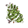
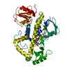
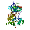
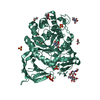
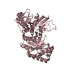
- PDB-4zw9: Crystal structure of human GLUT3 bound to D-glucose in the outwar... -
+ Open data

- Basic information
| Entry | Database: PDB / ID: 4zw9 | ||||||
|---|---|---|---|---|---|---|---|
| Title | Crystal structure of human GLUT3 bound to D-glucose in the outward-occluded conformation at 1.5 angstrom | ||||||
 Components Components | Solute carrier family 2, facilitated glucose transporter member 3 | ||||||
 Keywords Keywords | TRANSPORT PROTEIN / transporter | ||||||
| Function / homology |  Function and homology information Function and homology informationgalactose transmembrane transporter activity / galactose transmembrane transport / dehydroascorbic acid transmembrane transporter activity / dehydroascorbic acid transport / Vitamin C (ascorbate) metabolism / L-ascorbic acid metabolic process / Cellular hexose transport / D-glucose import across plasma membrane / D-glucose transmembrane transporter activity / D-glucose transmembrane transport ...galactose transmembrane transporter activity / galactose transmembrane transport / dehydroascorbic acid transmembrane transporter activity / dehydroascorbic acid transport / Vitamin C (ascorbate) metabolism / L-ascorbic acid metabolic process / Cellular hexose transport / D-glucose import across plasma membrane / D-glucose transmembrane transporter activity / D-glucose transmembrane transport / D-glucose binding / aggresome / : / tertiary granule membrane / ficolin-1-rich granule membrane / specific granule membrane / transport across blood-brain barrier / MECP2 regulates neuronal receptors and channels / cell projection / secretory granule membrane / carbohydrate metabolic process / perikaryon / Neutrophil degranulation / extracellular exosome / membrane / plasma membrane Similarity search - Function | ||||||
| Biological species |  Homo sapiens (human) Homo sapiens (human) | ||||||
| Method |  X-RAY DIFFRACTION / SYNCHROTRON / MOLECULAR REPLACEMENT / Resolution: 1.502 Å X-RAY DIFFRACTION / SYNCHROTRON / MOLECULAR REPLACEMENT / Resolution: 1.502 Å | ||||||
 Authors Authors | Deng, D. / Sun, P.C. / Yan, C.Y. / Yan, N. | ||||||
 Citation Citation | Journal: Nature / Year: 2015 Title: Molecular basis of ligand recognition and transport by glucose transporters Authors: Deng, D. / Sun, P.C. / Yan, C.Y. / Ke, M. / Jiang, X. / Xiong, L. / Ren, W. / Hirata, K. / Yamamoto, M. / Fan, S. / Yan, N. | ||||||
| History |
|
- Structure visualization
| Structure viewer | Molecule: MolmilJmol/JSmol |
|---|
- Downloads & links
-Download
| PDBx/mmCIF format | 4zw9.cif.gz | 198.7 KB | Display | PDBx/mmCIF format |
|---|---|---|---|---|
| PDB format | pdb4zw9.ent.gz | 157.3 KB | Display | PDB format |
| PDBx/mmJSON format | 4zw9.json.gz | Tree view | PDBx/mmJSON format | |
| Others |  Other downloads Other downloads |
-Validation report
| Arichive directory | https://data.pdbj.org/pub/pdb/validation_reports/zw/4zw9ftp://data.pdbj.org/pub/pdb/validation_reports/zw/4zw9 | HTTPS FTP |
|---|
-Related structure data
| Related structure data | 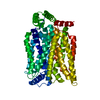 4zwbC  4zwcC  4pypS C: citing same article ( S: Starting model for refinement |
|---|---|
| Similar structure data |


-Links
 PDBj
PDBj


- Assembly
| Deposited unit | 
| ||||||||
|---|---|---|---|---|---|---|---|---|---|
| 1 |
| ||||||||
| Unit cell |
|
-Components
| #1: Protein | Mass: 56474.492 Da / Num. of mol.: 1 / Mutation: N43T Source method: isolated from a genetically manipulated source Source: (gene. exp.) Homo sapiens (human) / Gene: SLC2A3, GLUT3 / Production host:   Spodoptera frugiperda (fall armyworm) / References: UniProt: P11169 Spodoptera frugiperda (fall armyworm) / References: UniProt: P11169 | ||||||
|---|---|---|---|---|---|---|---|
| #2: Chemical |   Mass: 356.540 Da / Num. of mol.: 3 / Source method: obtained synthetically / Formula: C21H40O4 Mass: 356.540 Da / Num. of mol.: 3 / Source method: obtained synthetically / Formula: C21H40O4#3: Sugar | ChemComp-GLC / |   Type: D-saccharide, alpha linking / Mass: 180.156 Da / Num. of mol.: 1 Type: D-saccharide, alpha linking / Mass: 180.156 Da / Num. of mol.: 1Source method: isolated from a genetically manipulated source Formula: C6H12O6 #4: Sugar | ChemComp-BGC / |   Type: D-saccharide, beta linking / Mass: 180.156 Da / Num. of mol.: 1 Type: D-saccharide, beta linking / Mass: 180.156 Da / Num. of mol.: 1Source method: isolated from a genetically manipulated source Formula: C6H12O6 #5: Water | ChemComp-HOH / |  Mass: 18.015 Da / Num. of mol.: 101 / Source method: isolated from a natural source / Formula: H2O Mass: 18.015 Da / Num. of mol.: 101 / Source method: isolated from a natural source / Formula: H2O |
-Experimental details
-Experiment
| Experiment | Method: X-RAY DIFFRACTION |
|---|
- Sample preparation
| Crystal | Density Matthews: 2.53 Å3/Da / Density % sol: 51.43 % |
|---|---|
| Crystal grow | Temperature: 293 K / Method: lipidic cubic phase / pH: 6.8 Details: 28%(v/v) PEG400, 0.1M HEPES, 50 mM ammonium citrate |
-Data collection
| Diffraction | Mean temperature: 100 K |
|---|---|
| Diffraction source | Source: SYNCHROTRON / Site: SPring-8  / Beamline: BL32XU / Wavelength: 1 Å / Beamline: BL32XU / Wavelength: 1 Å |
| Detector | Type: RAYONIX MX225HE / Detector: CCD / Date: Dec 4, 2014 |
| Radiation | Protocol: SINGLE WAVELENGTH / Monochromatic (M) / Laue (L): M / Scattering type: x-ray |
| Radiation wavelength | Wavelength: 1 Å / Relative weight: 1 |
| Reflection | Resolution: 1.5→40 Å / Num. obs: 88120 / % possible obs: 98.8 % / Redundancy: 18 % / Rmerge(I) obs: 0.086 / Net I/σ(I): 12.7 |
| Reflection shell | Resolution: 1.5→1.55 Å / Redundancy: 2 % / Rmerge(I) obs: 0.578 / Mean I/σ(I) obs: 3.6 / % possible all: 91.8 |
- Processing
| Software | Name: PHENIX / Version: (phenix.refine: 1.9_1692) / Classification: refinement | |||||||||||||||||||||||||||||||||||||||||||||||||||||||||||||||||||||||||||||||||||||||||||||||||||||||||||||||||||||||||||||||||||||||||||||||||||||||||||||||||||||||||||||||||||||||||||||||||||||||||||||||||||||||||
|---|---|---|---|---|---|---|---|---|---|---|---|---|---|---|---|---|---|---|---|---|---|---|---|---|---|---|---|---|---|---|---|---|---|---|---|---|---|---|---|---|---|---|---|---|---|---|---|---|---|---|---|---|---|---|---|---|---|---|---|---|---|---|---|---|---|---|---|---|---|---|---|---|---|---|---|---|---|---|---|---|---|---|---|---|---|---|---|---|---|---|---|---|---|---|---|---|---|---|---|---|---|---|---|---|---|---|---|---|---|---|---|---|---|---|---|---|---|---|---|---|---|---|---|---|---|---|---|---|---|---|---|---|---|---|---|---|---|---|---|---|---|---|---|---|---|---|---|---|---|---|---|---|---|---|---|---|---|---|---|---|---|---|---|---|---|---|---|---|---|---|---|---|---|---|---|---|---|---|---|---|---|---|---|---|---|---|---|---|---|---|---|---|---|---|---|---|---|---|---|---|---|---|---|---|---|---|---|---|---|---|---|---|---|---|---|---|---|---|
| Refinement | Method to determine structure: MOLECULAR REPLACEMENT Starting model: 4pyp Resolution: 1.502→30.226 Å / SU ML: 0.12 / Cross valid method: FREE R-VALUE / σ(F): 1.36 / Phase error: 20.15 / Stereochemistry target values: ML
| |||||||||||||||||||||||||||||||||||||||||||||||||||||||||||||||||||||||||||||||||||||||||||||||||||||||||||||||||||||||||||||||||||||||||||||||||||||||||||||||||||||||||||||||||||||||||||||||||||||||||||||||||||||||||
| Solvent computation | Shrinkage radii: 0.9 Å / VDW probe radii: 1.11 Å / Solvent model: FLAT BULK SOLVENT MODEL | |||||||||||||||||||||||||||||||||||||||||||||||||||||||||||||||||||||||||||||||||||||||||||||||||||||||||||||||||||||||||||||||||||||||||||||||||||||||||||||||||||||||||||||||||||||||||||||||||||||||||||||||||||||||||
| Refinement step | Cycle: LAST / Resolution: 1.502→30.226 Å
| |||||||||||||||||||||||||||||||||||||||||||||||||||||||||||||||||||||||||||||||||||||||||||||||||||||||||||||||||||||||||||||||||||||||||||||||||||||||||||||||||||||||||||||||||||||||||||||||||||||||||||||||||||||||||
| Refine LS restraints |
| |||||||||||||||||||||||||||||||||||||||||||||||||||||||||||||||||||||||||||||||||||||||||||||||||||||||||||||||||||||||||||||||||||||||||||||||||||||||||||||||||||||||||||||||||||||||||||||||||||||||||||||||||||||||||
| LS refinement shell |
|
