[English] 日本語
Yorodumi
- PDB-4awi: Human Jnk1alpha kinase with 4-phenyl-7-azaindole IKK2 inhibitor. -

+
Open data


ID or keywords:

Loading...

-
Basic information

Entry
Database: PDB / ID: 4awi
TitleHuman Jnk1alpha kinase with 4-phenyl-7-azaindole IKK2 inhibitor.
ComponentsMITOGEN-ACTIVATED PROTEIN KINASE 8
KeywordsTRANSFERASE
Function / homology
Function and homology information


positive regulation of cell killing / JUN phosphorylation / Interleukin-38 signaling / Activation of BMF and translocation to mitochondria / basal dendrite / Activation of BIM and translocation to mitochondria / JUN kinase activity / WNT5:FZD7-mediated leishmania damping / positive regulation of cyclase activity / histone deacetylase regulator activity ...positive regulation of cell killing / JUN phosphorylation / Interleukin-38 signaling / Activation of BMF and translocation to mitochondria / basal dendrite / Activation of BIM and translocation to mitochondria / JUN kinase activity / WNT5:FZD7-mediated leishmania damping / positive regulation of cyclase activity / histone deacetylase regulator activity / positive regulation of NLRP3 inflammasome complex assembly / DSCAM interactions / NRAGE signals death through JNK / Activation of the AP-1 family of transcription factors / Fc-epsilon receptor signaling pathway / mitogen-activated protein kinase / regulation of macroautophagy / response to mechanical stimulus / response to UV / protein serine/threonine kinase binding / stress-activated MAPK cascade / JNK cascade / cellular response to cadmium ion / positive regulation of protein metabolic process / cellular response to amino acid starvation / NRIF signals cell death from the nucleus / JNK (c-Jun kinases) phosphorylation and activation mediated by activated human TAK1 / negative regulation of protein binding / FCERI mediated MAPK activation / peptidyl-threonine phosphorylation / regulation of circadian rhythm / histone deacetylase binding / cellular response to reactive oxygen species / cellular response to mechanical stimulus / regulation of protein localization / cellular senescence / Signaling by ALK fusions and activated point mutants / rhythmic process / Recruitment and ATM-mediated phosphorylation of repair and signaling proteins at DNA double strand breaks / cellular response to oxidative stress / cellular response to lipopolysaccharide / peptidyl-serine phosphorylation / protein phosphatase binding / Oxidative Stress Induced Senescence / response to oxidative stress / positive regulation of apoptotic process / axon / protein phosphorylation / protein serine kinase activity / protein serine/threonine kinase activity / synapse / positive regulation of gene expression / negative regulation of apoptotic process / enzyme binding / nucleoplasm / ATP binding / nucleus / cytoplasm / cytosol
Similarity search - Function
Mitogen-activated protein (MAP) kinase, JNK / Mitogen-activated protein (MAP) kinase, conserved site / MAP kinase signature. / Transferase(Phosphotransferase) domain 1 / Transferase(Phosphotransferase); domain 1 / Phosphorylase Kinase; domain 1 / Phosphorylase Kinase; domain 1 / Serine/threonine-protein kinase, active site / Serine/Threonine protein kinases active-site signature. / Protein kinase domain ...Mitogen-activated protein (MAP) kinase, JNK / Mitogen-activated protein (MAP) kinase, conserved site / MAP kinase signature. / Transferase(Phosphotransferase) domain 1 / Transferase(Phosphotransferase); domain 1 / Phosphorylase Kinase; domain 1 / Phosphorylase Kinase; domain 1 / Serine/threonine-protein kinase, active site / Serine/Threonine protein kinases active-site signature. / Protein kinase domain / Serine/Threonine protein kinases, catalytic domain / Protein kinase domain profile. / Protein kinase domain / Protein kinase-like domain superfamily / 2-Layer Sandwich / Orthogonal Bundle / Mainly Alpha / Alpha Beta
Similarity search - Domain/homology
Chem-AQ2 / Mitogen-activated protein kinase 8
Similarity search - Component
Biological speciesHOMO SAPIENS (human)
MethodX-RAY DIFFRACTION / SYNCHROTRON / MOLECULAR REPLACEMENT / Resolution: 1.91 Å
AuthorsChung, C. / Vicentini, G. / Liddle, J. / Bamborough, P.
CitationJournal: Bioorg.Med.Chem.Lett. / Year: 2012
Title: 4-Phenyl-7-Azaindoles as Potent, Selective and Bioavailable Ikk2 Inhibitors Demonstrating Good in Vivo Efficacy.
Authors: Liddle, J. / Bamborough, P. / Barker, M.D. / Campos, S. / Chung, C. / Cousins, R.P.C. / Faulder, P. / Heathcote, M.L. / Hobbs, H. / Holmes, D.S. / Ioannou, C. / Ramirez-Molina, C. / Morse, M. ...Authors: Liddle, J. / Bamborough, P. / Barker, M.D. / Campos, S. / Chung, C. / Cousins, R.P.C. / Faulder, P. / Heathcote, M.L. / Hobbs, H. / Holmes, D.S. / Ioannou, C. / Ramirez-Molina, C. / Morse, M.A. / Osborn, R. / Payne, J.J. / Pritchard, J.M. / Rumsey, W.L. / Tape, D.T. / Vicentini, G. / Whitworth, C. / Williamson, R.A.
History
DepositionJun 3, 2012Deposition site: PDBE / Processing site: PDBE
Revision 1.0May 1, 2013Provider: repository / Type: Initial release
Revision 1.1Jul 30, 2014Group: Database references
Revision 1.2May 8, 2024Group: Data collection / Database references ...Data collection / Database references / Derived calculations / Other
Category: chem_comp_atom / chem_comp_bond ...chem_comp_atom / chem_comp_bond / database_2 / pdbx_database_status / struct_site
Item: _database_2.pdbx_DOI / _database_2.pdbx_database_accession ..._database_2.pdbx_DOI / _database_2.pdbx_database_accession / _pdbx_database_status.status_code_sf / _struct_site.pdbx_auth_asym_id / _struct_site.pdbx_auth_comp_id / _struct_site.pdbx_auth_seq_id

-
Structure visualization

Structure viewerMolecule:
MolmilJmol/JSmol

Downloads & links

-
Assembly

Deposited unit
A: MITOGEN-ACTIVATED PROTEIN KINASE 8
hetero molecules


Theoretical massNumber of molelcules
Total (without water)43,1484
Polymers42,5081
Non-polymers6403
Water5,152286
1


  • Idetical with deposited unit
  • defined by author&software
TypeNameSymmetry operationNumber
identity operation1_555x,y,z1
MethodPISA
Unit cell
Length a, b, c (Å)50.438, 72.747, 110.155
Angle α, β, γ (deg.)90.00, 90.00, 90.00
Int Tables number19
Space group name H-MP212121

-
Components

#1: Protein MITOGEN-ACTIVATED PROTEIN KINASE 8 / MAP KINASE 8 / MAPK 8 / JNK-46 / STRESS-ACTIVATED PROTEIN KINASE 1C / SAPK1C / STRESS-ACTIVATED ...MAP KINASE 8 / MAPK 8 / JNK-46 / STRESS-ACTIVATED PROTEIN KINASE 1C / SAPK1C / STRESS-ACTIVATED PROTEIN KINASE JNK1 / C- JUN N-TERMINAL KINASE 1


Mass: 42508.191 Da / Num. of mol.: 1 / Fragment: RESIDUES 1-364
Source method: isolated from a genetically manipulated source
Source: (gene. exp.) HOMO SAPIENS (human) / Production host: ESCHERICHIA COLI (E. coli)
References: UniProt: P45983, mitogen-activated protein kinase
#2: Chemical ChemComp-AQ2 / N-(1,1-dioxidotetrahydro-2H-thiopyran-4-yl)-4-[2-(1-methylethyl)-1H-pyrrolo[2,3-b]pyridin-4-yl]benzenesulfonamide


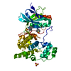
Mass: 447.571 Da / Num. of mol.: 1 / Source method: obtained synthetically / Formula: C21H25N3O4S2
#3: Chemical ChemComp-SO4 / SULFATE ION


Mass: 96.063 Da / Num. of mol.: 2 / Source method: obtained synthetically / Formula: SO4
#4: Water ChemComp-HOH / water


Mass: 18.015 Da / Num. of mol.: 286 / Source method: isolated from a natural source / Formula: H2O

-
Experimental details

-
Experiment

ExperimentMethod: X-RAY DIFFRACTION / Number of used crystals: 1

-
Sample preparation

CrystalDensity Matthews: 2.38 Å3/Da / Density % sol: 48.26 % / Description: NONE
Crystal growpH: 6.5
Details: 0.05 M AMMONIUM SULFATE, 0.05 M BIS-TRIS PH 6.5, 30% V/V PENTAERYTHRITOL (15/4 EO/OH)

-
Data collection

DiffractionMean temperature: 100 K
Diffraction sourceSource: SYNCHROTRON / Site: ESRF / Type: ESRF / Wavelength: 0.931
DetectorType: ADSC CCD / Detector: CCD / Date: Jul 1, 2006
RadiationProtocol: SINGLE WAVELENGTH / Monochromatic (M) / Laue (L): M / Scattering type: x-ray
Radiation wavelengthWavelength: 0.931 Å / Relative weight: 1
ReflectionResolution: 1.91→29.3 Å / Num. obs: 27663 / % possible obs: 89.3 % / Observed criterion σ(I): 2 / Redundancy: 2.8 % / Rmerge(I) obs: 0.06 / Net I/σ(I): 10.2
Reflection shellResolution: 1.91→2 Å / Redundancy: 2.6 % / Rmerge(I) obs: 0.39 / Mean I/σ(I) obs: 2.3 / % possible all: 86.4

-
Processing

Software
NameVersionClassification
REFMAC5.6.0117refinement
MOSFLMdata reduction
SCALAdata scaling
RefinementMethod to determine structure: MOLECULAR REPLACEMENT / Resolution: 1.91→28.5 Å / Cor.coef. Fo:Fc: 0.954 / Cor.coef. Fo:Fc free: 0.935 / SU B: 6.817 / SU ML: 0.105 / Cross valid method: THROUGHOUT / ESU R: 0.186 / ESU R Free: 0.157 / Stereochemistry target values: MAXIMUM LIKELIHOOD / Details: HYDROGENS HAVE BEEN ADDED IN THE RIDING POSITIONS.
RfactorNum. reflection% reflectionSelection details
Rfree0.21941 1391 5 %RANDOM
Rwork0.18601 ---
obs0.18769 26266 86.49 %-
Solvent computationIon probe radii: 0.8 Å / Shrinkage radii: 0.8 Å / VDW probe radii: 1.2 Å / Solvent model: BABINET MODEL WITH MASK
Displacement parametersBiso mean: 29.267 Å2
Baniso -1Baniso -2Baniso -3
1-1.91 Å20 Å20 Å2
2---0.63 Å20 Å2
3----1.28 Å2
Refinement stepCycle: LAST / Resolution: 1.91→28.5 Å
ProteinNucleic acidLigandSolventTotal
Num. atoms2832 0 40 286 3158
Refine LS restraints
Refine-IDTypeDev idealDev ideal targetNumber
X-RAY DIFFRACTIONr_bond_refined_d0.0070.0193001
X-RAY DIFFRACTIONr_bond_other_d0.0060.022040
X-RAY DIFFRACTIONr_angle_refined_deg1.0121.974085
X-RAY DIFFRACTIONr_angle_other_deg0.83635005
X-RAY DIFFRACTIONr_dihedral_angle_1_deg5.1395370
X-RAY DIFFRACTIONr_dihedral_angle_2_deg33.42624.632136
X-RAY DIFFRACTIONr_dihedral_angle_3_deg13.57615540
X-RAY DIFFRACTIONr_dihedral_angle_4_deg15.461515
X-RAY DIFFRACTIONr_chiral_restr0.060.2448
X-RAY DIFFRACTIONr_gen_planes_refined0.0040.0213297
X-RAY DIFFRACTIONr_gen_planes_other0.0010.02585
X-RAY DIFFRACTIONr_nbd_refined
X-RAY DIFFRACTIONr_nbd_other
X-RAY DIFFRACTIONr_nbtor_refined
X-RAY DIFFRACTIONr_nbtor_other
X-RAY DIFFRACTIONr_xyhbond_nbd_refined
X-RAY DIFFRACTIONr_xyhbond_nbd_other
X-RAY DIFFRACTIONr_metal_ion_refined
X-RAY DIFFRACTIONr_metal_ion_other
X-RAY DIFFRACTIONr_symmetry_vdw_refined
X-RAY DIFFRACTIONr_symmetry_vdw_other
X-RAY DIFFRACTIONr_symmetry_hbond_refined
X-RAY DIFFRACTIONr_symmetry_hbond_other
X-RAY DIFFRACTIONr_symmetry_metal_ion_refined
X-RAY DIFFRACTIONr_symmetry_metal_ion_other
X-RAY DIFFRACTIONr_mcbond_it
X-RAY DIFFRACTIONr_mcbond_other
X-RAY DIFFRACTIONr_mcangle_it
X-RAY DIFFRACTIONr_mcangle_other
X-RAY DIFFRACTIONr_scbond_it
X-RAY DIFFRACTIONr_scbond_other
X-RAY DIFFRACTIONr_scangle_it
X-RAY DIFFRACTIONr_scangle_other
X-RAY DIFFRACTIONr_long_range_B_refined
X-RAY DIFFRACTIONr_long_range_B_other
X-RAY DIFFRACTIONr_rigid_bond_restr
X-RAY DIFFRACTIONr_sphericity_free
X-RAY DIFFRACTIONr_sphericity_bonded
LS refinement shellResolution: 1.914→1.964 Å / Total num. of bins used: 20
RfactorNum. reflection% reflection
Rfree0.284 92 -
Rwork0.269 1537 -
obs--72.79 %
Refinement TLS params.Method: refined / Origin x: 16.0622 Å / Origin y: 15.9303 Å / Origin z: 23.8644 Å
111213212223313233
T0.0104 Å2-0.0132 Å2-0.0083 Å2-0.0267 Å20.0051 Å2--0.0203 Å2
L0.0621 °2-0.0717 °20.0891 °2-0.1003 °2-0.0329 °2--0.4102 °2
S-0.0153 Å °0.0117 Å °0.0128 Å °0.0237 Å °-0.0158 Å °-0.0133 Å °-0.0021 Å °0.0009 Å °0.0311 Å °

+
About Yorodumi

-
News

-
Feb 9, 2022. New format data for meta-information of EMDB entries

New format data for meta-information of EMDB entries

  • Version 3 of the EMDB header file is now the official format.
  • The previous official version 1.9 will be removed from the archive.

Related info.:EMDB header

External links:wwPDB to switch to version 3 of the EMDB data model

-
Aug 12, 2020. Covid-19 info

Covid-19 info

URL: https://pdbjlvh1.pdbj.org/emnavi/covid19.php

New page: Covid-19 featured information page in EM Navigator.

Related info.:Covid-19 info / Mar 5, 2020. Novel coronavirus structure data

+
Mar 5, 2020. Novel coronavirus structure data

Novel coronavirus structure data

Related info.:Yorodumi Speices / Aug 12, 2020. Covid-19 info

External links:COVID-19 featured content - PDBj / Molecule of the Month (242):Coronavirus Proteases

+
Jan 31, 2019. EMDB accession codes are about to change! (news from PDBe EMDB page)

EMDB accession codes are about to change! (news from PDBe EMDB page)

  • The allocation of 4 digits for EMDB accession codes will soon come to an end. Whilst these codes will remain in use, new EMDB accession codes will include an additional digit and will expand incrementally as the available range of codes is exhausted. The current 4-digit format prefixed with “EMD-” (i.e. EMD-XXXX) will advance to a 5-digit format (i.e. EMD-XXXXX), and so on. It is currently estimated that the 4-digit codes will be depleted around Spring 2019, at which point the 5-digit format will come into force.
  • The EM Navigator/Yorodumi systems omit the EMD- prefix.

Related info.:Q: What is EMD? / ID/Accession-code notation in Yorodumi/EM Navigator

External links:EMDB Accession Codes are Changing Soon! / Contact to PDBj

+
Jul 12, 2017. Major update of PDB

Major update of PDB

  • wwPDB released updated PDB data conforming to the new PDBx/mmCIF dictionary.
  • This is a major update changing the version number from 4 to 5, and with Remediation, in which all the entries are updated.
  • In this update, many items about electron microscopy experimental information are reorganized (e.g. em_software).
  • Now, EM Navigator and Yorodumi are based on the updated data.

External links:wwPDB Remediation / Enriched Model Files Conforming to OneDep Data Standards Now Available in the PDB FTP Archive

-
Yorodumi

Thousand views of thousand structures

  • Yorodumi is a browser for structure data from EMDB, PDB, SASBDB, etc.
  • This page is also the successor to EM Navigator detail page, and also detail information page/front-end page for Omokage search.
  • The word "yorodu" (or yorozu) is an old Japanese word meaning "ten thousand". "mi" (miru) is to see.

Related info.:EMDB / PDB / SASBDB / Comparison of 3 databanks / Yorodumi Search / Aug 31, 2016. New EM Navigator & Yorodumi / Yorodumi Papers / Jmol/JSmol / Function and homology information / Changes in new EM Navigator and Yorodumi

Read more