 Movie
Movie Controller

[English] 日本語
 Yorodumi
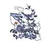
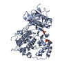
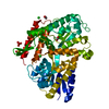
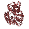
- PDB-2gmx: Selective Aminopyridine-Based C-Jun N-terminal Kinase inhibitors ... -
+ Open data

- Basic information
| Entry | Database: PDB / ID: 2gmx | ||||||
|---|---|---|---|---|---|---|---|
| Title | Selective Aminopyridine-Based C-Jun N-terminal Kinase inhibitors with cellular activity | ||||||
 Components Components |
| ||||||
 Keywords Keywords | TRANSCRIPTION / JNK1 / c-jun N-Terminal Kinase / Protein Kinase Jnk1 inhibitors / Aminopyridine-Based c-Jun N-Terminal Kinase Inhibitors | ||||||
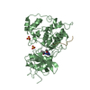
| Function / homology |  Function and homology information Function and homology informationdentate gyrus mossy fiber / JUN phosphorylation / positive regulation of cell killing / regulation of CD8-positive, alpha-beta T cell proliferation / Activation of BMF and translocation to mitochondria / Interleukin-38 signaling / basal dendrite / negative regulation of JUN kinase activity / JUN kinase activity / Activation of BIM and translocation to mitochondria ...dentate gyrus mossy fiber / JUN phosphorylation / positive regulation of cell killing / regulation of CD8-positive, alpha-beta T cell proliferation / Activation of BMF and translocation to mitochondria / Interleukin-38 signaling / basal dendrite / negative regulation of JUN kinase activity / JUN kinase activity / Activation of BIM and translocation to mitochondria / positive regulation of protein localization to mitochondrion / WNT5:FZD7-mediated leishmania damping / MAP kinase scaffold activity / JUN kinase binding / positive regulation of cyclase activity / histone deacetylase regulator activity / mitogen-activated protein kinase kinase binding / mitogen-activated protein kinase kinase kinase binding / regulation of JNK cascade / peptidyl-threonine phosphorylation / NRAGE signals death through JNK / Activation of the AP-1 family of transcription factors / Fc-epsilon receptor signaling pathway / dendritic growth cone / negative regulation of intrinsic apoptotic signaling pathway / protein kinase inhibitor activity / positive regulation of NLRP3 inflammasome complex assembly / positive regulation of protein metabolic process / mitogen-activated protein kinase / regulation of macroautophagy / negative regulation of protein binding / kinesin binding / response to mechanical stimulus / axonal growth cone / stress-activated MAPK cascade / response to UV / JNK cascade / vesicle-mediated transport / energy homeostasis / NRIF signals cell death from the nucleus / peptidyl-serine phosphorylation / cellular response to amino acid starvation / negative regulation of autophagy / JNK (c-Jun kinases) phosphorylation and activation mediated by activated human TAK1 / integrin-mediated signaling pathway / protein serine/threonine kinase binding / FCERI mediated MAPK activation / cellular response to reactive oxygen species / cellular response to mechanical stimulus / regulation of circadian rhythm / positive regulation of JNK cascade / autophagy / mitochondrial membrane / histone deacetylase binding / cellular senescence / Signaling by ALK fusions and activated point mutants / rhythmic process / MAPK cascade / regulation of protein localization / Recruitment and ATM-mediated phosphorylation of repair and signaling proteins at DNA double strand breaks / cellular response to lipopolysaccharide / cellular response to oxidative stress / response to oxidative stress / protein phosphatase binding / Oxidative Stress Induced Senescence / protein phosphorylation / positive regulation of apoptotic process / protein serine kinase activity / axon / protein serine/threonine kinase activity / neuronal cell body / positive regulation of gene expression / regulation of DNA-templated transcription / synapse / endoplasmic reticulum membrane / negative regulation of apoptotic process / perinuclear region of cytoplasm / enzyme binding / nucleoplasm / ATP binding / nucleus / plasma membrane / cytoplasm / cytosol Similarity search - Function | ||||||
| Biological species |  Homo sapiens (human) Homo sapiens (human) | ||||||
| Method |  X-RAY DIFFRACTION / SYNCHROTRON / MOLECULAR REPLACEMENT / Resolution: 3.5 Å X-RAY DIFFRACTION / SYNCHROTRON / MOLECULAR REPLACEMENT / Resolution: 3.5 Å | ||||||
 Authors Authors | Abad-Zapatero, C. | ||||||
 Citation Citation | Journal: J.Med.Chem. / Year: 2006 Title: Aminopyridine-Based c-Jun N-Terminal Kinase Inhibitors with Cellular Activity and Minimal Cross-Kinase Activity. Authors: Szczepankiewicz, B.G. / Kosogof, C. / Nelson, L.T. / Liu, G. / Liu, B. / Zhao, H. / Serby, M.D. / Xin, Z. / Liu, M. / Gum, R.J. / Haasch, D.L. / Wang, S. / Clampit, J.E. / Johnson, E.F. / ...Authors: Szczepankiewicz, B.G. / Kosogof, C. / Nelson, L.T. / Liu, G. / Liu, B. / Zhao, H. / Serby, M.D. / Xin, Z. / Liu, M. / Gum, R.J. / Haasch, D.L. / Wang, S. / Clampit, J.E. / Johnson, E.F. / Lubben, T.H. / Stashko, M.A. / Olejniczak, E.T. / Sun, C. / Dorwin, S.A. / Haskins, K. / Abad-Zapatero, C. / Fry, E.H. / Hutchins, C.W. / Sham, H.L. / Rondinone, C.M. / Trevillyan, J.M. #1: Journal: Bioorg.Med.Chem.Lett. / Year: 2006Title: Synthesis and SAR of 1,9-dihydro-9-hydroxypyrazolo[3,4-b]quinolin-4-ones as novel, selective c-Jun N-terminal kinase inhibitors. Authors: Liu, M. / Xin, Z. / Clampit, J.E. / Wang, S. / Gum, R.J. / Haasch, D.L. / Trevillyan, J.M. / Abad-Zapatero, C. / Fry, E.H. / Sham, H.L. / Liu, G. | ||||||
| History |
| ||||||
| Remark 999 | SEQUENCE THE NATIVE, UNMUTATED SEQUENCE IS THE SAME AS THE P45983-2 ISOFORM. THE INTRODUCED ...SEQUENCE THE NATIVE, UNMUTATED SEQUENCE IS THE SAME AS THE P45983-2 ISOFORM. THE INTRODUCED MUTATIONS (THR183>GLU, TYR185>GLU) ARE INTENDED TO MIMIC THE ACTIVATED FORM OF THE KINASE UPON PHOSPHORYLATION OF THOSE TWO RESIDUES. |
- Structure visualization
| Structure viewer | Molecule: MolmilJmol/JSmol |
|---|
- Downloads & links
-Download
| PDBx/mmCIF format | 2gmx.cif.gz | 149.3 KB | Display | PDBx/mmCIF format |
|---|---|---|---|---|
| PDB format | pdb2gmx.ent.gz | 117.8 KB | Display | PDB format |
| PDBx/mmJSON format | 2gmx.json.gz | Tree view | PDBx/mmJSON format | |
| Others |  Other downloads Other downloads |
-Validation report
| Arichive directory | https://data.pdbj.org/pub/pdb/validation_reports/gm/2gmxftp://data.pdbj.org/pub/pdb/validation_reports/gm/2gmx | HTTPS FTP |
|---|
-Related structure data
| Related structure data |  2g01S S: Starting model for refinement |
|---|---|
| Similar structure data |


-Links
 PDBj
PDBj


- Assembly
| Deposited unit | 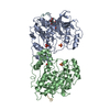
| ||||||||
|---|---|---|---|---|---|---|---|---|---|
| 1 | 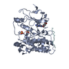
| ||||||||
| 2 | 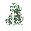
| ||||||||
| 3 |
| ||||||||
| Unit cell |
|
-Components
| #1: Protein | Mass: 42919.559 Da / Num. of mol.: 2 / Mutation: T183E,Y185E Source method: isolated from a genetically manipulated source Source: (gene. exp.) Homo sapiens (human) / Gene: MAPK8, JNK1, PRKM8 / Production host:  References: UniProt: P45983, mitogen-activated protein kinase #2: Protein/peptide | Mass: 1345.612 Da / Num. of mol.: 2 / Fragment: PepJIP1 peptide / Source method: obtained synthetically Details: THE SEQUENCE IS FOUND NATURALLY IN HOMO SAPIENS (HUMAN) References: UniProt: Q9UQF2 #3: Chemical | ChemComp-SO4 /   Mass: 96.063 Da / Num. of mol.: 4 / Source method: obtained synthetically / Formula: SO4 Mass: 96.063 Da / Num. of mol.: 4 / Source method: obtained synthetically / Formula: SO4#4: Chemical |   Mass: 435.272 Da / Num. of mol.: 2 / Source method: obtained synthetically / Formula: C18H19BrN4O4 Mass: 435.272 Da / Num. of mol.: 2 / Source method: obtained synthetically / Formula: C18H19BrN4O4 |
|---|
-Experimental details
-Experiment
| Experiment | Method: X-RAY DIFFRACTION / Number of used crystals: 1 |
|---|
- Sample preparation
| Crystal | Density Matthews: 4.4 Å3/Da / Density % sol: 72.02 % |
|---|---|
| Crystal grow | Temperature: 277 K / Method: vapor diffusion, hanging drop / pH: 6.2 Details: Protein was preincubated with the JIP1 peptide at a 5x molar excess. Protein concentration 9-12.6 mg/mL. Hanging drops consisted of 2uL protein plus 2 uL well solution. Well solution:2.8-3.1 ...Details: Protein was preincubated with the JIP1 peptide at a 5x molar excess. Protein concentration 9-12.6 mg/mL. Hanging drops consisted of 2uL protein plus 2 uL well solution. Well solution:2.8-3.1 M Ammonium Sulfate, 10-14% glycerol. For Co-crystallization experiment with the compound, the compound was dissolved in DMSO at 100 mM concentration. Allow to incubate for at least an hour on ice. Solution was spun for 5 minutes at 2000g prior to setting up for crystallization, pH 6.2, VAPOR DIFFUSION, HANGING DROP, temperature 277.0K |
-Data collection
| Diffraction | Mean temperature: 110 K |
|---|---|
| Diffraction source | Source: SYNCHROTRON / Site: APS  / Beamline: 17-ID / Wavelength: 1 Å / Beamline: 17-ID / Wavelength: 1 Å |
| Detector | Type: ADSC QUANTUM 210 / Detector: CCD / Date: Oct 18, 2004 |
| Radiation | Protocol: SINGLE WAVELENGTH / Monochromatic (M) / Laue (L): M / Scattering type: x-ray |
| Radiation wavelength | Wavelength: 1 Å / Relative weight: 1 |
| Reflection | Resolution: 3.5→20 Å / Num. all: 19198 / Num. obs: 17550 / % possible obs: 87.7 % / Observed criterion σ(F): 1 / Observed criterion σ(I): 1 / Redundancy: 5.6 % / Biso Wilson estimate: 21.5 Å2 / Rmerge(I) obs: 0.078 / Rsym value: 0.078 / Net I/σ(I): 21 |
| Reflection shell | Resolution: 3.5→3.62 Å / Redundancy: 5.8 % / Rmerge(I) obs: 0.385 / Mean I/σ(I) obs: 4.62 / Num. unique all: 1942 / Rsym value: 0.385 / % possible all: 99.3 |
- Processing
| Software |
| ||||||||||||||||||||||||||||||||||||||||||||||||||||||||||||
|---|---|---|---|---|---|---|---|---|---|---|---|---|---|---|---|---|---|---|---|---|---|---|---|---|---|---|---|---|---|---|---|---|---|---|---|---|---|---|---|---|---|---|---|---|---|---|---|---|---|---|---|---|---|---|---|---|---|---|---|---|---|
| Refinement | Method to determine structure: MOLECULAR REPLACEMENT Starting model: 2g01 Resolution: 3.5→19.91 Å / Rfactor Rfree error: 0.009 / Data cutoff high absF: 337281.36 / Data cutoff low absF: 0 / Isotropic thermal model: GROUP / Cross valid method: THROUGHOUT / σ(F): 2 / Stereochemistry target values: Engh & Huber
| ||||||||||||||||||||||||||||||||||||||||||||||||||||||||||||
| Solvent computation | Solvent model: FLAT MODEL / Bsol: 10 Å2 / ksol: 0.202359 e/Å3 | ||||||||||||||||||||||||||||||||||||||||||||||||||||||||||||
| Displacement parameters | Biso mean: 56.4 Å2
| ||||||||||||||||||||||||||||||||||||||||||||||||||||||||||||
| Refine analyze |
| ||||||||||||||||||||||||||||||||||||||||||||||||||||||||||||
| Refinement step | Cycle: LAST / Resolution: 3.5→19.91 Å
| ||||||||||||||||||||||||||||||||||||||||||||||||||||||||||||
| Refine LS restraints |
| ||||||||||||||||||||||||||||||||||||||||||||||||||||||||||||
| LS refinement shell | Resolution: 3.5→3.62 Å / Rfactor Rfree error: 0.04 / Total num. of bins used: 10
| ||||||||||||||||||||||||||||||||||||||||||||||||||||||||||||
| Xplor file |
|
