 Movie
Movie Controller

[English] 日本語
 Yorodumi
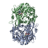
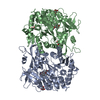
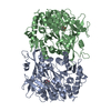
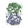
- PDB-4rmn: Crystal structure of a benzoate coenzyme A ligase with 2-Thiophen... -
+ Open data

- Basic information
| Entry | Database: PDB / ID: 4rmn | ||||||
|---|---|---|---|---|---|---|---|
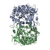
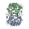
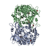
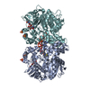
| Title | Crystal structure of a benzoate coenzyme A ligase with 2-Thiophene Carboxylic acid | ||||||
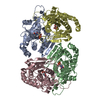
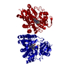
 Components Components | Benzoate-coenzyme A ligase | ||||||
 Keywords Keywords | LIGASE / Substrate Specificity / Kinetics | ||||||
| Function / homology |  Function and homology information Function and homology informationbenzoate-CoA ligase / benzoate-CoA ligase activity / acid-thiol ligase activity / CoA-ligase activity / secondary metabolite biosynthetic process / ATP binding Similarity search - Function | ||||||
| Biological species |  Rhodopseudomonas palustris (phototrophic) Rhodopseudomonas palustris (phototrophic) | ||||||
| Method |  X-RAY DIFFRACTION / SYNCHROTRON / MOLECULAR REPLACEMENT / Resolution: 1.72 Å X-RAY DIFFRACTION / SYNCHROTRON / MOLECULAR REPLACEMENT / Resolution: 1.72 Å | ||||||
 Authors Authors | Strom, S. / Nosrati, M. / Thornburg, C. / Walker, K.D. / Geiger, J.H. | ||||||
 Citation Citation | Journal: Biochemistry / Year: 2015 Title: Kinetically and Crystallographically Guided Mutations of a Benzoate CoA Ligase (BadA) Elucidate Mechanism and Expand Substrate Permissivity. Authors: Thornburg, C.K. / Wortas-Strom, S. / Nosrati, M. / Geiger, J.H. / Walker, K.D. | ||||||
| History |
|
- Structure visualization
| Structure viewer | Molecule: MolmilJmol/JSmol |
|---|
- Downloads & links
-Download
| PDBx/mmCIF format | 4rmn.cif.gz | 220.3 KB | Display | PDBx/mmCIF format |
|---|---|---|---|---|
| PDB format | pdb4rmn.ent.gz | 175 KB | Display | PDB format |
| PDBx/mmJSON format | 4rmn.json.gz | Tree view | PDBx/mmJSON format | |
| Others |  Other downloads Other downloads |
-Validation report
| Arichive directory | https://data.pdbj.org/pub/pdb/validation_reports/rm/4rmnftp://data.pdbj.org/pub/pdb/validation_reports/rm/4rmn | HTTPS FTP |
|---|
-Related structure data
| Related structure data |  4eatC  4rlfC  4rlqC  4rm2C  4rm3C  4zjzC  2v7bS C: citing same article ( S: Starting model for refinement |
|---|---|
| Similar structure data |
-Links
 PDBj
PDBj
- Assembly
| Deposited unit | 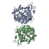
| ||||||||
|---|---|---|---|---|---|---|---|---|---|
| 1 | 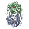
| ||||||||
| 2 | 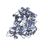
| ||||||||
| 3 | 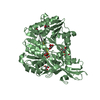
| ||||||||
| Unit cell |
|
-Components
| #1: Protein | Mass: 56792.246 Da / Num. of mol.: 2 Source method: isolated from a genetically manipulated source Source: (gene. exp.) Rhodopseudomonas palustris (phototrophic)Gene: badA / Plasmid: pET28a / Production host: #2: Chemical | ChemComp-C21 / 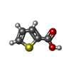  Mass: 128.149 Da / Num. of mol.: 4 / Source method: obtained synthetically / Formula: C5H4O2S Mass: 128.149 Da / Num. of mol.: 4 / Source method: obtained synthetically / Formula: C5H4O2S#3: Chemical | ChemComp-GOL /   Mass: 92.094 Da / Num. of mol.: 7 / Source method: obtained synthetically / Formula: C3H8O3 Mass: 92.094 Da / Num. of mol.: 7 / Source method: obtained synthetically / Formula: C3H8O3#4: Water | ChemComp-HOH / |  Mass: 18.015 Da / Num. of mol.: 572 / Source method: isolated from a natural source / Formula: H2O Mass: 18.015 Da / Num. of mol.: 572 / Source method: isolated from a natural source / Formula: H2O |
|---|
-Experimental details
-Experiment
| Experiment | Method: X-RAY DIFFRACTION / Number of used crystals: 1 |
|---|
- Sample preparation
| Crystal | Density Matthews: 2.26 Å3/Da / Density % sol: 45.54 % |
|---|---|
| Crystal grow | Temperature: 298 K / Method: vapor diffusion, hanging drop / pH: 7 Details: 15 % PEG 3350, 0.1 M Tris pH 7.0, VAPOR DIFFUSION, HANGING DROP, temperature 298K |
-Data collection
| Diffraction | Mean temperature: 100 K |
|---|---|
| Diffraction source | Source: SYNCHROTRON / Site: APS  / Beamline: 21-ID-G / Wavelength: 0.97872 Å / Beamline: 21-ID-G / Wavelength: 0.97872 Å |
| Detector | Type: MARMOSAIC 300 mm CCD / Detector: CCD / Date: Feb 12, 2011 |
| Radiation | Protocol: SINGLE WAVELENGTH / Monochromatic (M) / Laue (L): M / Scattering type: x-ray |
| Radiation wavelength | Wavelength: 0.97872 Å / Relative weight: 1 |
| Reflection | Resolution: 1.72→39.58 Å / Num. obs: 99134 / % possible obs: 97.57 % / Redundancy: 4.4 % / Biso Wilson estimate: 25.4 Å2 / Rmerge(I) obs: 0.097 / Rsym value: 0.079 / Net I/σ(I): 21.84 |
| Reflection shell | Resolution: 1.72→1.76 Å / Redundancy: 3 % / Rmerge(I) obs: 0.472 / Mean I/σ(I) obs: 1.97 / Rsym value: 0.417 / % possible all: 80 |
- Processing
| Software |
| |||||||||||||||||||||||||||||||||||||||||||||||||||||||||||||||||||||||||||||||||||||||||||||||||||||||||
|---|---|---|---|---|---|---|---|---|---|---|---|---|---|---|---|---|---|---|---|---|---|---|---|---|---|---|---|---|---|---|---|---|---|---|---|---|---|---|---|---|---|---|---|---|---|---|---|---|---|---|---|---|---|---|---|---|---|---|---|---|---|---|---|---|---|---|---|---|---|---|---|---|---|---|---|---|---|---|---|---|---|---|---|---|---|---|---|---|---|---|---|---|---|---|---|---|---|---|---|---|---|---|---|---|---|---|
| Refinement | Method to determine structure: MOLECULAR REPLACEMENT Starting model: 2V7B Resolution: 1.72→39.58 Å / Cor.coef. Fo:Fc: 0.971 / Cor.coef. Fo:Fc free: 0.958 / SU B: 2.364 / SU ML: 0.074 / Cross valid method: THROUGHOUT / ESU R: 0.101 / ESU R Free: 0.101 / Stereochemistry target values: MAXIMUM LIKELIHOOD / Details: HYDROGENS HAVE BEEN ADDED IN THE RIDING POSITIONS
| |||||||||||||||||||||||||||||||||||||||||||||||||||||||||||||||||||||||||||||||||||||||||||||||||||||||||
| Solvent computation | Ion probe radii: 0.8 Å / Shrinkage radii: 0.8 Å / VDW probe radii: 1.2 Å / Solvent model: MASK | |||||||||||||||||||||||||||||||||||||||||||||||||||||||||||||||||||||||||||||||||||||||||||||||||||||||||
| Displacement parameters | Biso mean: 25.67 Å2
| |||||||||||||||||||||||||||||||||||||||||||||||||||||||||||||||||||||||||||||||||||||||||||||||||||||||||
| Refinement step | Cycle: LAST / Resolution: 1.72→39.58 Å
| |||||||||||||||||||||||||||||||||||||||||||||||||||||||||||||||||||||||||||||||||||||||||||||||||||||||||
| Refine LS restraints |
| |||||||||||||||||||||||||||||||||||||||||||||||||||||||||||||||||||||||||||||||||||||||||||||||||||||||||
| LS refinement shell | Resolution: 1.72→1.765 Å / Total num. of bins used: 20
|
