 Movie
Movie Controller

[English] 日本語
 Yorodumi
- PDB-2h7c: Crystal structure of human carboxylesterase in complex with Coenzyme A -
+ Open data

- Basic information
| Entry | Database: PDB / ID: 2h7c | |||||||||
|---|---|---|---|---|---|---|---|---|---|---|
| Title | Crystal structure of human carboxylesterase in complex with Coenzyme A | |||||||||
 Components Components | Liver carboxylesterase 1 | |||||||||
 Keywords Keywords | HYDROLASE / enzyme / esterase / cholesteryl esterase | |||||||||
| Function / homology |  Function and homology information Function and homology informationcholesterol ester hydrolysis involved in cholesterol transport / methylumbelliferyl-acetate deacetylase / methylumbelliferyl-acetate deacetylase activity / regulation of bile acid secretion / sterol esterase / sterol ester esterase activity / medium-chain fatty acid metabolic process / carboxylesterase / Physiological factors / carboxylesterase activity ...cholesterol ester hydrolysis involved in cholesterol transport / methylumbelliferyl-acetate deacetylase / methylumbelliferyl-acetate deacetylase activity / regulation of bile acid secretion / sterol esterase / sterol ester esterase activity / medium-chain fatty acid metabolic process / carboxylesterase / Physiological factors / carboxylesterase activity / regulation of bile acid biosynthetic process / cellular response to cholesterol / reverse cholesterol transport / positive regulation of cholesterol metabolic process / Phase I - Functionalization of compounds / carboxylic ester hydrolase activity / cholesterol biosynthetic process / Aspirin ADME / negative regulation of cholesterol storage / positive regulation of cholesterol efflux / cellular response to low-density lipoprotein particle stimulus / cholesterol metabolic process / Metabolism of Angiotensinogen to Angiotensins / lipid catabolic process / epithelial cell differentiation / lipid droplet / cholesterol homeostasis / response to toxic substance / endoplasmic reticulum lumen / endoplasmic reticulum / cytoplasm / cytosol Similarity search - Function | |||||||||
| Biological species |  Homo sapiens (human) Homo sapiens (human) | |||||||||
| Method |  X-RAY DIFFRACTION / SYNCHROTRON / MOLECULAR REPLACEMENT / Resolution: 2 Å X-RAY DIFFRACTION / SYNCHROTRON / MOLECULAR REPLACEMENT / Resolution: 2 Å | |||||||||
 Authors Authors | Bencharit, S. / Edwards, C.C. / Morton, C.L. / Howard-Williams, E.L. / Potter, P.M. / Redinbo, M.R. | |||||||||
 Citation Citation | Journal: J.Mol.Biol. / Year: 2006 Title: Multisite promiscuity in the processing of endogenous substrates by human carboxylesterase 1 Authors: Bencharit, S. / Edwards, C.C. / Morton, C.L. / Howard-Williams, E.L. / Kuhn, P. / Potter, P.M. / Redinbo, M.R. | |||||||||
| History |
|
- Structure visualization
| Structure viewer | Molecule: MolmilJmol/JSmol |
|---|
- Downloads & links
-Download
| PDBx/mmCIF format | 2h7c.cif.gz | 697.9 KB | Display | PDBx/mmCIF format |
|---|---|---|---|---|
| PDB format | pdb2h7c.ent.gz | 560.1 KB | Display | PDB format |
| PDBx/mmJSON format | 2h7c.json.gz | Tree view | PDBx/mmJSON format | |
| Others |  Other downloads Other downloads |
-Validation report
| Arichive directory | https://data.pdbj.org/pub/pdb/validation_reports/h7/2h7cftp://data.pdbj.org/pub/pdb/validation_reports/h7/2h7c | HTTPS FTP |
|---|
-Related structure data
| Related structure data | 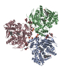 2dqyC  2dqzC 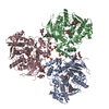 2dr0C  1mx1S S: Starting model for refinement C: citing same article ( |
|---|---|
| Similar structure data |


-Links
 PDBj
PDBj


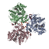
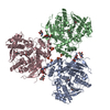
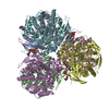
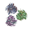
- Assembly
| Deposited unit | 
| ||||||||
|---|---|---|---|---|---|---|---|---|---|
| 1 |
| ||||||||
| 2 | 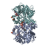
| ||||||||
| 3 | 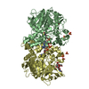
| ||||||||
| 4 | 
| ||||||||
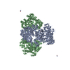
| Unit cell |
|
-Components
-Protein , 1 types, 6 molecules ABCDEF
| #1: Protein | Mass: 59787.527 Da / Num. of mol.: 6 / Fragment: RESIDUES 19-561 Source method: isolated from a genetically manipulated source Source: (gene. exp.) Homo sapiens (human) / Cell line (production host): sf9 / Production host:   Spodoptera frugiperda (fall armyworm) / References: UniProt: P23141, carboxylesterase Spodoptera frugiperda (fall armyworm) / References: UniProt: P23141, carboxylesterase |
|---|
-Sugars , 3 types, 8 molecules 

| #2: Polysaccharide | Source method: isolated from a genetically manipulated source #5: Sugar | ChemComp-NAG /  Type: D-saccharide, beta linking / Mass: 221.208 Da / Num. of mol.: 4 Type: D-saccharide, beta linking / Mass: 221.208 Da / Num. of mol.: 4Source method: isolated from a genetically manipulated source Formula: C8H15NO6 #6: Sugar |  Type: D-saccharide, alpha linking / Mass: 309.270 Da / Num. of mol.: 2 Type: D-saccharide, alpha linking / Mass: 309.270 Da / Num. of mol.: 2Source method: isolated from a genetically manipulated source Formula: C11H19NO9 |
|---|
-Non-polymers , 3 types, 2733 molecules 


| #3: Chemical | ChemComp-SO4 /  Mass: 96.063 Da / Num. of mol.: 12 / Source method: obtained synthetically / Formula: SO4 Mass: 96.063 Da / Num. of mol.: 12 / Source method: obtained synthetically / Formula: SO4#4: Chemical | ChemComp-COA /  Mass: 767.534 Da / Num. of mol.: 6 / Source method: obtained synthetically / Formula: C21H36N7O16P3S Mass: 767.534 Da / Num. of mol.: 6 / Source method: obtained synthetically / Formula: C21H36N7O16P3S#7: Water | ChemComp-HOH / | Mass: 18.015 Da / Num. of mol.: 2715 / Source method: isolated from a natural source / Formula: H2O |
|---|
-Details
| Has protein modification | Y |
|---|
-Experimental details
-Experiment
| Experiment | Method: X-RAY DIFFRACTION / Number of used crystals: 1 |
|---|
- Sample preparation
| Crystal | Density Matthews: 2.51 Å3/Da / Density % sol: 51 % |
|---|---|
| Crystal grow | Temperature: 298 K / Method: vapor diffusion, sitting drop / pH: 5.6 Details: 8% PEG 3350, 0.4M Li2SO4, 0.1M NaCl, 0.1M LiCl, 0.1M citrate (pH 5.5), 5% glycerol, pH 5.6, VAPOR DIFFUSION, SITTING DROP, temperature 298K |
-Data collection
| Diffraction source | Source: SYNCHROTRON / Site: SSRL  / Beamline: BL11-1 / Beamline: BL11-1 |
|---|---|
| Detector | Date: Mar 1, 2001 |
| Radiation | Protocol: SINGLE WAVELENGTH / Monochromatic (M) / Laue (L): M / Scattering type: x-ray |
| Radiation wavelength | Relative weight: 1 |
| Reflection | Resolution: 2→30 Å / Num. obs: 233023 / % possible obs: 97.1 % / Observed criterion σ(F): 2 / Observed criterion σ(I): 2 / Redundancy: 7.7 % / Biso Wilson estimate: 15.5 Å2 / Rsym value: 0.082 / Net I/σ(I): 12.7 |
| Reflection shell | Resolution: 2→2.13 Å / Mean I/σ(I) obs: 2.9 / Rsym value: 0.335 / % possible all: 88.1 |
- Processing
| Software |
| ||||||||||||||||||||||||||||||||||||
|---|---|---|---|---|---|---|---|---|---|---|---|---|---|---|---|---|---|---|---|---|---|---|---|---|---|---|---|---|---|---|---|---|---|---|---|---|---|
| Refinement | Method to determine structure: MOLECULAR REPLACEMENT Starting model: PDB ENTRY 1MX1 Resolution: 2→29.1 Å / Rfactor Rfree error: 0.002 / Data cutoff high absF: 3374696.62 / Data cutoff low absF: 0 / Isotropic thermal model: RESTRAINED / Cross valid method: THROUGHOUT / σ(F): 0 / Stereochemistry target values: Engh & Huber
| ||||||||||||||||||||||||||||||||||||
| Solvent computation | Solvent model: FLAT MODEL / Bsol: 48.2004 Å2 / ksol: 0.362937 e/Å3 | ||||||||||||||||||||||||||||||||||||
| Displacement parameters | Biso mean: 28.4 Å2
| ||||||||||||||||||||||||||||||||||||
| Refine analyze |
| ||||||||||||||||||||||||||||||||||||
| Refinement step | Cycle: LAST / Resolution: 2→29.1 Å
| ||||||||||||||||||||||||||||||||||||
| Refine LS restraints |
| ||||||||||||||||||||||||||||||||||||
| LS refinement shell | Resolution: 2→2.13 Å / Rfactor Rfree error: 0.007 / Total num. of bins used: 6
| ||||||||||||||||||||||||||||||||||||
| Xplor file |
|
