 Movie
Movie Controller

[English] 日本語
 Yorodumi
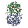
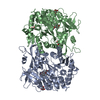
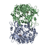
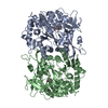
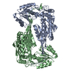
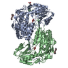
- PDB-6m2u: The crystal structure of benzoate coenzyme A ligase double mutant... -
+ Open data

- Basic information
| Entry | Database: PDB / ID: 6m2u | ||||||
|---|---|---|---|---|---|---|---|
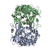
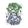
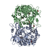
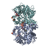
| Title | The crystal structure of benzoate coenzyme A ligase double mutant (H333A/I334A) in complex with 2-chloro-1,3-thiazole-5-carboxylate-AMP | ||||||
 Components Components | Benzoate-coenzyme A ligase | ||||||
 Keywords Keywords | LIGASE / Benzoate / coenzyme / complex | ||||||
| Function / homology |  Function and homology information Function and homology informationacid-thiol ligase activity / CoA-ligase activity / secondary metabolite biosynthetic process / ATP binding Similarity search - Function | ||||||
| Biological species |  Rhodopseudomonas palustris (phototrophic) Rhodopseudomonas palustris (phototrophic) | ||||||
| Method |  X-RAY DIFFRACTION / SYNCHROTRON / MOLECULAR REPLACEMENT / Resolution: 1.709 Å X-RAY DIFFRACTION / SYNCHROTRON / MOLECULAR REPLACEMENT / Resolution: 1.709 Å | ||||||
 Authors Authors | Li, T.L. / Adhikari, K. | ||||||
 Citation Citation | Journal: Biomolecules / Year: 2020 Title: Chemoenzymatic Synthesis and Biological Evaluation for Bioactive Molecules Derived from Bacterial Benzoyl Coenzyme A Ligase and Plant Type III Polyketide Synthase. Authors: Adhikari, K. / Lo, I.W. / Chen, C.L. / Wang, Y.L. / Lin, K.H. / Zadeh, S.M. / Rattinam, R. / Li, Y.S. / Wu, C.J. / Li, T.L. | ||||||
| History |
|
- Structure visualization
| Structure viewer | Molecule: MolmilJmol/JSmol |
|---|
- Downloads & links
-Download
| PDBx/mmCIF format | 6m2u.cif.gz | 243.5 KB | Display | PDBx/mmCIF format |
|---|---|---|---|---|
| PDB format | pdb6m2u.ent.gz | 188.1 KB | Display | PDB format |
| PDBx/mmJSON format | 6m2u.json.gz | Tree view | PDBx/mmJSON format | |
| Others |  Other downloads Other downloads |
-Validation report
| Arichive directory | https://data.pdbj.org/pub/pdb/validation_reports/m2/6m2uftp://data.pdbj.org/pub/pdb/validation_reports/m2/6m2u | HTTPS FTP |
|---|
-Related structure data
| Related structure data |  6m2oSC S: Starting model for refinement C: citing same article ( |
|---|---|
| Similar structure data |
-Links
 PDBj
PDBj


- Assembly
| Deposited unit | 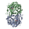
| ||||||||
|---|---|---|---|---|---|---|---|---|---|
| 1 |
| ||||||||
| Unit cell |
|
-Components
| #1: Protein | Mass: 56167.500 Da / Num. of mol.: 2 / Mutation: T83A, H333A, I334A, G341D Source method: isolated from a genetically manipulated source Source: (gene. exp.) Rhodopseudomonas palustris (phototrophic)Gene: badA / Production host: #2: Chemical |   Mass: 347.221 Da / Num. of mol.: 2 Mass: 347.221 Da / Num. of mol.: 2Source method: isolated from a genetically manipulated source Formula: C10H14N5O7P / Comment: AMP*YM #3: Chemical | 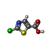  Mass: 163.582 Da / Num. of mol.: 2 Mass: 163.582 Da / Num. of mol.: 2Source method: isolated from a genetically manipulated source Formula: C4H2ClNO2S #4: Water | ChemComp-HOH / |  Mass: 18.015 Da / Num. of mol.: 1307 / Source method: isolated from a natural source / Formula: H2O Mass: 18.015 Da / Num. of mol.: 1307 / Source method: isolated from a natural source / Formula: H2OHas ligand of interest | Y | |
|---|
-Experimental details
-Experiment
| Experiment | Method: X-RAY DIFFRACTION / Number of used crystals: 1 |
|---|
- Sample preparation
| Crystal | Density Matthews: 2.25 Å3/Da / Density % sol: 45.27 % |
|---|---|
| Crystal grow | Temperature: 293 K / Method: vapor diffusion, hanging drop / pH: 6.5 / Details: PEG 4000, 0.1 M sodium chloride, 0.2 M MES |
-Data collection
| Diffraction | Mean temperature: 200 K / Serial crystal experiment: N |
|---|---|
| Diffraction source | Source: SYNCHROTRON / Site: NSRRC  / Beamline: BL15A1 / Wavelength: 1 Å / Beamline: BL15A1 / Wavelength: 1 Å |
| Detector | Type: RAYONIX MX300HE / Detector: CCD / Date: Jun 19, 2019 |
| Radiation | Protocol: SINGLE WAVELENGTH / Monochromatic (M) / Laue (L): M / Scattering type: x-ray |
| Radiation wavelength | Wavelength: 1 Å / Relative weight: 1 |
| Reflection | Resolution: 1.709→29.816 Å / Num. obs: 105468 / % possible obs: 99.9 % / Redundancy: 6.7 % / CC1/2: 0.97 / Net I/σ(I): 33.03 |
| Reflection shell | Resolution: 1.709→1.709 Å / Num. unique obs: 105468 / CC1/2: 0.887 |
- Processing
| Software |
| |||||||||||||||||||||||||||||||||||||||||||||||||||||||||||||||||||||||||||||||||||||||||||||||||||||||||
|---|---|---|---|---|---|---|---|---|---|---|---|---|---|---|---|---|---|---|---|---|---|---|---|---|---|---|---|---|---|---|---|---|---|---|---|---|---|---|---|---|---|---|---|---|---|---|---|---|---|---|---|---|---|---|---|---|---|---|---|---|---|---|---|---|---|---|---|---|---|---|---|---|---|---|---|---|---|---|---|---|---|---|---|---|---|---|---|---|---|---|---|---|---|---|---|---|---|---|---|---|---|---|---|---|---|---|
| Refinement | Method to determine structure: MOLECULAR REPLACEMENT Starting model: 6M2O Resolution: 1.709→29.816 Å / SU ML: 0.18 / Cross valid method: NONE / σ(F): 1.34 / Phase error: 20.41 / Stereochemistry target values: ML
| |||||||||||||||||||||||||||||||||||||||||||||||||||||||||||||||||||||||||||||||||||||||||||||||||||||||||
| Solvent computation | Shrinkage radii: 0.9 Å / VDW probe radii: 1.11 Å / Solvent model: FLAT BULK SOLVENT MODEL | |||||||||||||||||||||||||||||||||||||||||||||||||||||||||||||||||||||||||||||||||||||||||||||||||||||||||
| Refinement step | Cycle: LAST / Resolution: 1.709→29.816 Å
| |||||||||||||||||||||||||||||||||||||||||||||||||||||||||||||||||||||||||||||||||||||||||||||||||||||||||
| Refine LS restraints |
| |||||||||||||||||||||||||||||||||||||||||||||||||||||||||||||||||||||||||||||||||||||||||||||||||||||||||
| LS refinement shell |
|
