 Movie
Movie Controller

[English] 日本語
 Yorodumi
- PDB-5too: Crystal structure of alkaline phosphatase PafA T79S, N100A, K162A... -
+ Open data

- Basic information
| Entry | Database: PDB / ID: 5too | ||||||
|---|---|---|---|---|---|---|---|
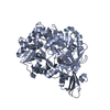
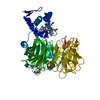
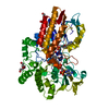
| Title | Crystal structure of alkaline phosphatase PafA T79S, N100A, K162A, R164A mutant | ||||||
 Components Components | Alkaline phosphatase PafA | ||||||
 Keywords Keywords | HYDROLASE / Alkaline Phosphatase / Phosphomonoesterase / PafA / Weak phosphate binder / Zinc bimetallo core | ||||||
| Function / homology |  Function and homology information Function and homology informationcarbohydrate phosphatase activity / glucose-1-phosphatase activity / glucose-6-phosphatase activity / sugar-phosphatase activity / alkaline phosphatase / alkaline phosphatase activity / periplasmic space / zinc ion binding / metal ion binding Similarity search - Function | ||||||
| Biological species |  Elizabethkingia meningoseptica (bacteria) Elizabethkingia meningoseptica (bacteria) | ||||||
| Method |  X-RAY DIFFRACTION / SYNCHROTRON / MOLECULAR REPLACEMENT / molecular replacement / Resolution: 2.031 Å X-RAY DIFFRACTION / SYNCHROTRON / MOLECULAR REPLACEMENT / molecular replacement / Resolution: 2.031 Å | ||||||
 Authors Authors | Lyubimov, A.Y. / Sunden, F. / AlSadhan, I. / Herschlag, D. | ||||||
| Funding support |  United States, 1items United States, 1items
| ||||||
 Citation Citation | Journal: J. Biol. Chem. / Year: 2017 Title: Differential catalytic promiscuity of the alkaline phosphatase superfamily bimetallo core reveals mechanistic features underlying enzyme evolution. Authors: Sunden, F. / AlSadhan, I. / Lyubimov, A. / Doukov, T. / Swan, J. / Herschlag, D. | ||||||
| History |
|
- Structure visualization
| Structure viewer | Molecule: MolmilJmol/JSmol |
|---|
- Downloads & links
-Download
| PDBx/mmCIF format | 5too.cif.gz | 204 KB | Display | PDBx/mmCIF format |
|---|---|---|---|---|
| PDB format | pdb5too.ent.gz | 162.1 KB | Display | PDB format |
| PDBx/mmJSON format | 5too.json.gz | Tree view | PDBx/mmJSON format | |
| Others |  Other downloads Other downloads |
-Validation report
| Arichive directory | https://data.pdbj.org/pub/pdb/validation_reports/to/5tooftp://data.pdbj.org/pub/pdb/validation_reports/to/5too | HTTPS FTP |
|---|
-Related structure data
| Related structure data | 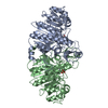 5tpqC  5tj3S S: Starting model for refinement C: citing same article ( |
|---|---|
| Similar structure data |


-Links
 PDBj
PDBj
- Assembly
| Deposited unit | 
| ||||||||
|---|---|---|---|---|---|---|---|---|---|
| 1 |
| ||||||||
| Unit cell |
|
-Components
| #1: Protein | Mass: 61120.406 Da / Num. of mol.: 1 / Fragment: UNP residues 21-546 Source method: isolated from a genetically manipulated source Source: (gene. exp.) Elizabethkingia meningoseptica (bacteria)Gene: pafA / Production host: | ||||
|---|---|---|---|---|---|
| #2: Chemical | ChemComp-ZN /   Mass: 65.409 Da / Num. of mol.: 4 / Source method: obtained synthetically / Formula: Zn Mass: 65.409 Da / Num. of mol.: 4 / Source method: obtained synthetically / Formula: Zn#3: Chemical | ChemComp-CL / |   Mass: 35.453 Da / Num. of mol.: 1 / Source method: obtained synthetically / Formula: Cl Mass: 35.453 Da / Num. of mol.: 1 / Source method: obtained synthetically / Formula: Cl#4: Water | ChemComp-HOH / |  Mass: 18.015 Da / Num. of mol.: 216 / Source method: isolated from a natural source / Formula: H2O Mass: 18.015 Da / Num. of mol.: 216 / Source method: isolated from a natural source / Formula: H2O |
-Experimental details
-Experiment
| Experiment | Method: X-RAY DIFFRACTION / Number of used crystals: 1 |
|---|
- Sample preparation
| Crystal | Density Matthews: 1.95 Å3/Da / Density % sol: 32.9 % |
|---|---|
| Crystal grow | Temperature: 291 K / Method: vapor diffusion, hanging drop / pH: 4.4 Details: 22% PEG3350, 0.1 M sodium acetate, pH 4.4, 0.2 M ammonium sulfate |
-Data collection
| Diffraction | Mean temperature: 100 K |
|---|---|
| Diffraction source | Source: SYNCHROTRON / Site: SSRL / Beamline: BL11-1 / Wavelength: 0.9795 Å |
| Detector | Type: DECTRIS PILATUS 6M / Detector: PIXEL / Date: Mar 31, 2016 |
| Radiation | Monochromator: Si(111) Side scattering bent cube-root I-beam single crystal Protocol: SINGLE WAVELENGTH / Monochromatic (M) / Laue (L): M / Scattering type: x-ray |
| Radiation wavelength | Wavelength: 0.9795 Å / Relative weight: 1 |
| Reflection | Resolution: 2.03→41.01 Å / Num. obs: 28568 / % possible obs: 99.9 % / Redundancy: 8.5 % / Biso Wilson estimate: 30.4 Å2 / CC1/2: 0.995 / Rmerge(I) obs: 0.168 / Net I/σ(I): 9.6 |
| Reflection shell | Resolution: 2.03→2.08 Å / Redundancy: 8.8 % / Rmerge(I) obs: 1.518 / CC1/2: 0.66 / % possible all: 99.9 |
-Phasing
| Phasing | Method: molecular replacement | |||||||||
|---|---|---|---|---|---|---|---|---|---|---|
| Phasing MR |
|
- Processing
| Software |
| ||||||||||||||||||||||||||||||||||||||||||||||||||||||||||||||||||||||||
|---|---|---|---|---|---|---|---|---|---|---|---|---|---|---|---|---|---|---|---|---|---|---|---|---|---|---|---|---|---|---|---|---|---|---|---|---|---|---|---|---|---|---|---|---|---|---|---|---|---|---|---|---|---|---|---|---|---|---|---|---|---|---|---|---|---|---|---|---|---|---|---|---|---|
| Refinement | Method to determine structure: MOLECULAR REPLACEMENT Starting model: 5TJ3 Resolution: 2.031→41.009 Å / SU ML: 0.23 / Cross valid method: FREE R-VALUE / σ(F): 1.35 / Phase error: 22.01
| ||||||||||||||||||||||||||||||||||||||||||||||||||||||||||||||||||||||||
| Solvent computation | Shrinkage radii: 0.9 Å / VDW probe radii: 1.11 Å | ||||||||||||||||||||||||||||||||||||||||||||||||||||||||||||||||||||||||
| Displacement parameters | Biso max: 99.1 Å2 / Biso mean: 40.6973 Å2 / Biso min: 16.98 Å2 | ||||||||||||||||||||||||||||||||||||||||||||||||||||||||||||||||||||||||
| Refinement step | Cycle: final / Resolution: 2.031→41.009 Å
| ||||||||||||||||||||||||||||||||||||||||||||||||||||||||||||||||||||||||
| Refine LS restraints |
| ||||||||||||||||||||||||||||||||||||||||||||||||||||||||||||||||||||||||
| LS refinement shell | Refine-ID: X-RAY DIFFRACTION / Total num. of bins used: 11 / % reflection obs: 100 %
|
