 Movie
Movie Controller


+ Open data

- Basic information
| Entry | Database: PDB / ID: 1gso | ||||||
|---|---|---|---|---|---|---|---|
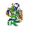
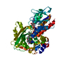
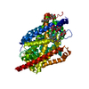
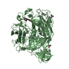
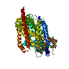
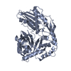
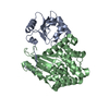
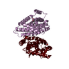
| Title | GLYCINAMIDE RIBONUCLEOTIDE SYNTHETASE (GAR-SYN) FROM E. COLI. | ||||||
 Components Components | PROTEIN (GLYCINAMIDE RIBONUCLEOTIDE SYNTHETASE) | ||||||
 Keywords Keywords | LIGASE / GAR-SYN / GLYCINAMIDE RIBONUCLEOTIDE SYNTHETASE / ATP-GRASP / PURINE DE NOVO BIOSYNTHETIC PATHWAY / SUBSTRATE CHANNELING | ||||||
| Function / homology |  Function and homology information Function and homology informationphosphoribosylamine-glycine ligase / purine nucleobase biosynthetic process / phosphoribosylamine-glycine ligase activity / purine nucleotide biosynthetic process / 'de novo' IMP biosynthetic process / DNA damage response / ATP hydrolysis activity / ATP binding / metal ion binding Similarity search - Function | ||||||
| Biological species |  | ||||||
| Method |  X-RAY DIFFRACTION / SYNCHROTRON / MAD / Resolution: 1.6 Å X-RAY DIFFRACTION / SYNCHROTRON / MAD / Resolution: 1.6 Å | ||||||
 Authors Authors | Wang, W. / Kappock, T.J. / Stubbe, J. / Ealick, S.E. | ||||||
 Citation Citation | Journal: Biochemistry / Year: 1998 Title: X-ray crystal structure of glycinamide ribonucleotide synthetase from Escherichia coli. Authors: Wang, W. / Kappock, T.J. / Stubbe, J. / Ealick, S.E. #1: Journal: Acta Crystallogr.,Sect.D / Year: 1999Title: Purification, Crystallization and Preliminary X-Ray Diffraction Data from Selenomethione Glycinamide Ribonucleotide Synthetase Authors: Weaver, T.M. / Wang, W. / Ealick, S.E. | ||||||
| History |
|
- Structure visualization
| Structure viewer | Molecule: MolmilJmol/JSmol |
|---|
- Downloads & links
-Download
| PDBx/mmCIF format | 1gso.cif.gz | 96.9 KB | Display | PDBx/mmCIF format |
|---|---|---|---|---|
| PDB format | pdb1gso.ent.gz | 74.5 KB | Display | PDB format |
| PDBx/mmJSON format | 1gso.json.gz | Tree view | PDBx/mmJSON format | |
| Others |  Other downloads Other downloads |
-Validation report
| Arichive directory | https://data.pdbj.org/pub/pdb/validation_reports/gs/1gsoftp://data.pdbj.org/pub/pdb/validation_reports/gs/1gso | HTTPS FTP |
|---|
-Related structure data
| Similar structure data |
|---|


-Links
 PDBj
PDBj


- Assembly
| Deposited unit | 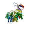
| ||||||||
|---|---|---|---|---|---|---|---|---|---|
| 1 |
| ||||||||
| Unit cell |
|
-Components
| #1: Protein | Mass: 46283.527 Da / Num. of mol.: 1 / Mutation: P294L / Source method: isolated from a natural source / Source: (natural) References: UniProt: P15640, phosphoribosylamine-glycine ligase |
|---|---|
| #2: Water | ChemComp-HOH /  Mass: 18.015 Da / Num. of mol.: 299 / Source method: isolated from a natural source / Formula: H2O Mass: 18.015 Da / Num. of mol.: 299 / Source method: isolated from a natural source / Formula: H2O |
-Experimental details
-Experiment
| Experiment | Method: X-RAY DIFFRACTION / Number of used crystals: 1 |
|---|
- Sample preparation
| Crystal | Density Matthews: 2.47 Å3/Da / Density % sol: 50 % | ||||||||||||||||||||
|---|---|---|---|---|---|---|---|---|---|---|---|---|---|---|---|---|---|---|---|---|---|
| Crystal grow | Method: vapor diffusion, hanging drop / pH: 6.3 Details: 0.1M MES PH6.3, 0.2M AMMONIUM SULFATE, 26% PEG 5K MONOMETHYLETHER, HANGING DROP VAPOR DEFFUSION, vapor diffusion - hanging drop | ||||||||||||||||||||
| Crystal | *PLUS Density % sol: 50 % | ||||||||||||||||||||
| Crystal grow | *PLUS Temperature: 18 ℃ / Method: vapor diffusion, hanging drop | ||||||||||||||||||||
| Components of the solutions | *PLUS
|
-Data collection
| Diffraction | Mean temperature: 93 K | |||||||||||||||
|---|---|---|---|---|---|---|---|---|---|---|---|---|---|---|---|---|
| Diffraction source | Source: SYNCHROTRON / Site: CHESS  / Beamline: F2 / Wavelength: 0.979419, 0.979104, 0.967642, 0.9190 / Beamline: F2 / Wavelength: 0.979419, 0.979104, 0.967642, 0.9190 | |||||||||||||||
| Detector | Type: ADSC / Detector: CCD / Date: Aug 1, 1997 | |||||||||||||||
| Radiation | Protocol: MAD / Monochromatic (M) / Laue (L): M / Scattering type: x-ray | |||||||||||||||
| Radiation wavelength |
| |||||||||||||||
| Reflection | Resolution: 1.6→20 Å / Num. obs: 58385 / % possible obs: 95.9 % / Redundancy: 5.2 % / Rsym value: 4.3 / Net I/σ(I): 10 | |||||||||||||||
| Reflection shell | Resolution: 1.6→1.69 Å / Redundancy: 4.1 % / Mean I/σ(I) obs: 4.1 / Rsym value: 17.4 / % possible all: 83.9 | |||||||||||||||
| Reflection | *PLUS Rmerge(I) obs: 0.043 | |||||||||||||||
| Reflection shell | *PLUS % possible obs: 83.9 % / Num. unique obs: 7256 / Rmerge(I) obs: 0.174 |
- Processing
| Software |
| ||||||||||||||||||||||||||||||||||||||||||||||||||||||||||||
|---|---|---|---|---|---|---|---|---|---|---|---|---|---|---|---|---|---|---|---|---|---|---|---|---|---|---|---|---|---|---|---|---|---|---|---|---|---|---|---|---|---|---|---|---|---|---|---|---|---|---|---|---|---|---|---|---|---|---|---|---|---|
| Refinement | Method to determine structure: MAD / Resolution: 1.6→10 Å / Data cutoff high absF: 1000000 / Data cutoff low absF: 0.001 / Cross valid method: THROUGHOUT / σ(F): 3
| ||||||||||||||||||||||||||||||||||||||||||||||||||||||||||||
| Displacement parameters | Biso mean: 22.1 Å2 | ||||||||||||||||||||||||||||||||||||||||||||||||||||||||||||
| Refine analyze | Luzzati coordinate error free: 0.24 Å | ||||||||||||||||||||||||||||||||||||||||||||||||||||||||||||
| Refinement step | Cycle: LAST / Resolution: 1.6→10 Å
| ||||||||||||||||||||||||||||||||||||||||||||||||||||||||||||
| Refine LS restraints |
| ||||||||||||||||||||||||||||||||||||||||||||||||||||||||||||
| LS refinement shell | Resolution: 1.6→1.67 Å / Total num. of bins used: 8
| ||||||||||||||||||||||||||||||||||||||||||||||||||||||||||||
| Xplor file |
| ||||||||||||||||||||||||||||||||||||||||||||||||||||||||||||
| Software | *PLUS Name: X-PLOR / Version: 3.851 / Classification: refinement | ||||||||||||||||||||||||||||||||||||||||||||||||||||||||||||
| Refinement | *PLUS Highest resolution: 1.6 Å / Lowest resolution: 10 Å / σ(F): 3 / % reflection Rfree: 10 % | ||||||||||||||||||||||||||||||||||||||||||||||||||||||||||||
| Solvent computation | *PLUS | ||||||||||||||||||||||||||||||||||||||||||||||||||||||||||||
| Displacement parameters | *PLUS Biso mean: 22.1 Å2 | ||||||||||||||||||||||||||||||||||||||||||||||||||||||||||||
| Refine LS restraints | *PLUS
| ||||||||||||||||||||||||||||||||||||||||||||||||||||||||||||
| LS refinement shell | *PLUS Highest resolution: 1.6 Å / Rfactor Rfree: 0.307 / % reflection Rfree: 10 % / Rfactor Rwork: 0.298 |
