[English] 日本語
Yorodumi
- PDB-1yru: Crystal Structure analysis of the adenylyl cyclaes catalytic doma... -

+
Open data


ID or keywords:

Loading...

-
Basic information

Entry
Database: PDB / ID: 1yru
Title
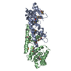
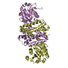
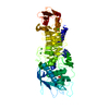
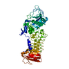
Crystal Structure analysis of the adenylyl cyclaes catalytic domain of adenylyl cyclase toxin of Bordetella pertussis in presence of c-terminal calmodulin and 1mM calcium chloride
Components
  • Bifunctional hemolysin-adenylate cyclase
  • Calmodulin
KeywordsLAYSE / TOXIN / CyaA / CaM / adenylyl Cyclase
Function / homology
Function and homology information


calcium- and calmodulin-responsive adenylate cyclase activity / hemolysis in another organism / : / : / positive regulation of cyclic-nucleotide phosphodiesterase activity / : / adenylate cyclase / negative regulation of peptidyl-threonine phosphorylation / establishment of protein localization to mitochondrial membrane / cAMP biosynthetic process ...calcium- and calmodulin-responsive adenylate cyclase activity / hemolysis in another organism / : / : / positive regulation of cyclic-nucleotide phosphodiesterase activity / : / adenylate cyclase / negative regulation of peptidyl-threonine phosphorylation / establishment of protein localization to mitochondrial membrane / cAMP biosynthetic process / type 3 metabotropic glutamate receptor binding / adenylate cyclase activity / CaM pathway / Cam-PDE 1 activation / Sodium/Calcium exchangers / Calmodulin induced events / Reduction of cytosolic Ca++ levels / Activation of Ca-permeable Kainate Receptor / CREB1 phosphorylation through the activation of CaMKII/CaMKK/CaMKIV cascasde / Loss of phosphorylation of MECP2 at T308 / CREB1 phosphorylation through the activation of Adenylate Cyclase / positive regulation of DNA binding / positive regulation of peptidyl-threonine phosphorylation / CaMK IV-mediated phosphorylation of CREB / PKA activation / negative regulation of high voltage-gated calcium channel activity / Glycogen breakdown (glycogenolysis) / positive regulation of ryanodine-sensitive calcium-release channel activity / CLEC7A (Dectin-1) induces NFAT activation / Activation of RAC1 downstream of NMDARs / negative regulation of calcium ion export across plasma membrane / organelle localization by membrane tethering / response to corticosterone / mitochondrion-endoplasmic reticulum membrane tethering / autophagosome membrane docking / nitric-oxide synthase binding / presynaptic endocytosis / regulation of cardiac muscle cell action potential / negative regulation of ryanodine-sensitive calcium-release channel activity / regulation of synaptic vesicle exocytosis / Synthesis of IP3 and IP4 in the cytosol / regulation of cell communication by electrical coupling involved in cardiac conduction / Phase 0 - rapid depolarisation / Negative regulation of NMDA receptor-mediated neuronal transmission / Unblocking of NMDA receptors, glutamate binding and activation / RHO GTPases activate PAKs / calcineurin-mediated signaling / Ion transport by P-type ATPases / positive regulation of protein autophosphorylation / Uptake and function of anthrax toxins / regulation of ryanodine-sensitive calcium-release channel activity / regulation of synaptic vesicle endocytosis / Long-term potentiation / Calcineurin activates NFAT / Regulation of MECP2 expression and activity / protein phosphatase activator activity / adenylate cyclase binding / DARPP-32 events / Smooth Muscle Contraction / catalytic complex / detection of calcium ion / positive regulation of protein serine/threonine kinase activity / regulation of cardiac muscle contraction / RHO GTPases activate IQGAPs / regulation of cardiac muscle contraction by regulation of the release of sequestered calcium ion / phosphatidylinositol 3-kinase binding / presynaptic cytosol / calcium channel inhibitor activity / cellular response to interferon-beta / Protein methylation / Activation of AMPK downstream of NMDARs / activation of adenylate cyclase activity / Ion homeostasis / regulation of release of sequestered calcium ion into cytosol by sarcoplasmic reticulum / eNOS activation / enzyme regulator activity / regulation of calcium-mediated signaling / Tetrahydrobiopterin (BH4) synthesis, recycling, salvage and regulation / titin binding / voltage-gated potassium channel complex / sperm midpiece / substantia nigra development / calcium channel complex / positive regulation of nitric-oxide synthase activity / calyx of Held / FCERI mediated Ca+2 mobilization / Ras activation upon Ca2+ influx through NMDA receptor / nitric-oxide synthase regulator activity / response to amphetamine / FCGR3A-mediated IL10 synthesis / adenylate cyclase activator activity / regulation of heart rate / Antigen activates B Cell Receptor (BCR) leading to generation of second messengers / protein serine/threonine kinase activator activity / VEGFR2 mediated cell proliferation / sarcomere / regulation of cytokinesis / VEGFR2 mediated vascular permeability / Translocation of SLC2A4 (GLUT4) to the plasma membrane / positive regulation of receptor signaling pathway via JAK-STAT
Similarity search - Function
DNA polymerase; domain 1 - #920 / Alpha-Beta Plaits - #1720 / Adenylylcyclase toxin fold / Anthrax toxin, edema factor, central domain / RTX, pore-forming domain / N-terminal domain in RTX protein / RTX toxin determinant A / Haemolysin-type calcium binding-related / Haemolysin-type calcium binding protein related domain / : ...DNA polymerase; domain 1 - #920 / Alpha-Beta Plaits - #1720 / Adenylylcyclase toxin fold / Anthrax toxin, edema factor, central domain / RTX, pore-forming domain / N-terminal domain in RTX protein / RTX toxin determinant A / Haemolysin-type calcium binding-related / Haemolysin-type calcium binding protein related domain / : / Anthrax toxin, edema factor, central / Anthrax toxin, edema factor, C-terminal / Anthrax toxin, edema factor, central domain superfamily / Anthrax toxin LF subunit / Hemolysin-type calcium-binding conserved site / Hemolysin-type calcium-binding region signature. / RTX calcium-binding nonapeptide repeat / RTX calcium-binding nonapeptide repeat (4 copies) / Serralysin-like metalloprotease, C-terminal / EF-hand / : / Recoverin; domain 1 / EF-hand domain pair / EF-hand, calcium binding motif / EF-Hand 1, calcium-binding site / EF-hand calcium-binding domain. / EF-hand calcium-binding domain profile. / DNA polymerase; domain 1 / EF-hand domain / EF-hand domain pair / Alpha-Beta Plaits / Alpha-Beta Complex / 2-Layer Sandwich / Orthogonal Bundle / Mainly Alpha / Alpha Beta
Similarity search - Domain/homology
Bifunctional hemolysin/adenylate cyclase / Calmodulin-1 / Bifunctional hemolysin/adenylate cyclase / Calmodulin-3
Similarity search - Component
Biological speciesBordetella pertussis (bacteria)
Homo sapiens (human)
MethodX-RAY DIFFRACTION / SYNCHROTRON / MOLECULAR REPLACEMENT / Resolution: 2.5 Å
AuthorsGuo, Q. / Shen, Y. / Tang, W.J.
CitationJournal: Embo J. / Year: 2005
Title: Structural basis for the interaction of Bordetella pertussis adenylyl cyclase toxin with calmodulin
Authors: Guo, Q. / Shen, Y. / Lee, Y.S. / Gibbs, C.S. / Mrksich, M. / Tang, W.J.
History
DepositionFeb 4, 2005Deposition site: RCSB / Processing site: RCSB
Revision 1.0Sep 27, 2005Provider: repository / Type: Initial release
Revision 1.1Apr 30, 2008Group: Version format compliance
Revision 1.2Jul 13, 2011Group: Version format compliance
Revision 1.3Feb 14, 2024Group: Data collection / Database references / Derived calculations
Category: chem_comp_atom / chem_comp_bond ...chem_comp_atom / chem_comp_bond / database_2 / pdbx_struct_conn_angle / struct_conn / struct_site
Item: _database_2.pdbx_DOI / _database_2.pdbx_database_accession ..._database_2.pdbx_DOI / _database_2.pdbx_database_accession / _pdbx_struct_conn_angle.ptnr1_auth_comp_id / _pdbx_struct_conn_angle.ptnr1_auth_seq_id / _pdbx_struct_conn_angle.ptnr1_label_asym_id / _pdbx_struct_conn_angle.ptnr1_label_atom_id / _pdbx_struct_conn_angle.ptnr1_label_comp_id / _pdbx_struct_conn_angle.ptnr1_label_seq_id / _pdbx_struct_conn_angle.ptnr2_auth_seq_id / _pdbx_struct_conn_angle.ptnr2_label_asym_id / _pdbx_struct_conn_angle.ptnr3_auth_comp_id / _pdbx_struct_conn_angle.ptnr3_auth_seq_id / _pdbx_struct_conn_angle.ptnr3_label_asym_id / _pdbx_struct_conn_angle.ptnr3_label_atom_id / _pdbx_struct_conn_angle.ptnr3_label_comp_id / _pdbx_struct_conn_angle.ptnr3_label_seq_id / _pdbx_struct_conn_angle.value / _struct_conn.pdbx_dist_value / _struct_conn.ptnr1_auth_comp_id / _struct_conn.ptnr1_auth_seq_id / _struct_conn.ptnr1_label_asym_id / _struct_conn.ptnr1_label_atom_id / _struct_conn.ptnr1_label_comp_id / _struct_conn.ptnr1_label_seq_id / _struct_conn.ptnr2_auth_comp_id / _struct_conn.ptnr2_auth_seq_id / _struct_conn.ptnr2_label_asym_id / _struct_conn.ptnr2_label_atom_id / _struct_conn.ptnr2_label_comp_id / _struct_conn.ptnr2_label_seq_id / _struct_site.pdbx_auth_asym_id / _struct_site.pdbx_auth_comp_id / _struct_site.pdbx_auth_seq_id

-
Structure visualization

Structure viewerMolecule:
MolmilJmol/JSmol

Downloads & links

-
Assembly

Deposited unit
A: Bifunctional hemolysin-adenylate cyclase
B: Calmodulin
hetero molecules


Theoretical massNumber of molelcules
Total (without water)48,0574
Polymers47,9772
Non-polymers802
Water1,26170
1


  • Idetical with deposited unit
  • defined by author&software
TypeNameSymmetry operationNumber
identity operation1_555x,y,z1
Buried area3400 Å2
ΔGint-28 kcal/mol
Surface area19490 Å2
MethodPISA
Unit cell
Length a, b, c (Å)79.441, 79.441, 139.181
Angle α, β, γ (deg.)90.00, 90.00, 90.00
Int Tables number92
Space group name H-MP41212

-
Components

#1: Protein Bifunctional hemolysin-adenylate cyclase / Cyclolysin / ACT / AC-HLY


Mass: 39431.859 Da / Num. of mol.: 1 / Fragment: Calmodulin-sensitive adenylate cyclase
Source method: isolated from a genetically manipulated source
Source: (gene. exp.) Bordetella pertussis (bacteria) / Gene: cya, cyaA / Plasmid: pEX / Species (production host): Escherichia coli / Production host: Escherichia coli BL21(DE3) (bacteria) / Strain (production host): BL21(DE3)
References: UniProt: P15318, UniProt: P0DKX7*PLUS, adenylate cyclase
#2: Protein Calmodulin / CaM / CALM / CAM1


Mass: 8545.383 Da / Num. of mol.: 1
Source method: isolated from a genetically manipulated source
Source: (gene. exp.) Homo sapiens (human) / Gene: CALM1, CALM2, CALM3 / Plasmid: pProEx / Production host: Escherichia coli (E. coli) / Strain (production host): BL21(DE3)pUBS520 / References: UniProt: P62158, UniProt: P0DP23*PLUS
#3: Chemical ChemComp-CA / CALCIUM ION


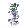
Mass: 40.078 Da / Num. of mol.: 2 / Source method: obtained synthetically / Formula: Ca
#4: Water ChemComp-HOH / water


Mass: 18.015 Da / Num. of mol.: 70 / Source method: isolated from a natural source / Formula: H2O

-
Experimental details

-
Experiment

ExperimentMethod: X-RAY DIFFRACTION / Number of used crystals: 1

-
Sample preparation

CrystalDensity Matthews: 2.4 Å3/Da / Density % sol: 56 %
Crystal growTemperature: 298 K / Method: vapor diffusion, hanging drop / pH: 6.2
Details: PEG2000, Tris, pH 6.2, VAPOR DIFFUSION, HANGING DROP, temperature 298K

-
Data collection

DiffractionMean temperature: 100 K
Diffraction sourceSource: SYNCHROTRON / Site: APS / Beamline: 19-ID / Wavelength: 0.97951 Å
DetectorType: CUSTOM-MADE / Detector: CCD / Date: Jul 17, 2004
RadiationProtocol: SINGLE WAVELENGTH / Monochromatic (M) / Laue (L): M / Scattering type: x-ray
Radiation wavelengthWavelength: 0.97951 Å / Relative weight: 1
ReflectionResolution: 2.5→30 Å / Num. all: 29428 / Num. obs: 27335 / % possible obs: 92.9 % / Observed criterion σ(F): 1 / Observed criterion σ(I): 1 / Biso Wilson estimate: 25.5 Å2
Reflection shellResolution: 2.5→2.68 Å / % possible all: 85

-
Processing

Software
NameVersionClassification
CNS1.1refinement
HKL-2000data reduction
SCALEPACKdata scaling
CNSphasing
RefinementMethod to determine structure: MOLECULAR REPLACEMENT / Resolution: 2.5→30 Å / Rfactor Rfree error: 0.008 / Data cutoff high absF: 451487.42 / Data cutoff low absF: 0 / Isotropic thermal model: RESTRAINED / Cross valid method: THROUGHOUT / σ(F): 0
RfactorNum. reflection% reflectionSelection details
Rfree0.308 1319 4.8 %RANDOM
Rwork0.231 ---
all0.24 ---
obs0.231 27335 92.9 %-
Solvent computationSolvent model: FLAT MODEL / Bsol: 20.2769 Å2 / ksol: 0.294173 e/Å3
Displacement parametersBiso mean: 34.7 Å2
Baniso -1Baniso -2Baniso -3
1-4.53 Å20 Å20 Å2
2--4.53 Å20 Å2
3----9.06 Å2
Refine analyze
FreeObs
Luzzati coordinate error0.47 Å0.32 Å
Luzzati d res low-5 Å
Luzzati sigma a0.39 Å0.28 Å
Refinement stepCycle: LAST / Resolution: 2.5→30 Å
ProteinNucleic acidLigandSolventTotal
Num. atoms3235 0 2 70 3307
Refine LS restraints
Refine-IDTypeDev ideal
X-RAY DIFFRACTIONc_bond_d0.022
X-RAY DIFFRACTIONc_bond_d_na
X-RAY DIFFRACTIONc_bond_d_prot
X-RAY DIFFRACTIONc_angle_d
X-RAY DIFFRACTIONc_angle_d_na
X-RAY DIFFRACTIONc_angle_d_prot
X-RAY DIFFRACTIONc_angle_deg2.4
X-RAY DIFFRACTIONc_angle_deg_na
X-RAY DIFFRACTIONc_angle_deg_prot
X-RAY DIFFRACTIONc_dihedral_angle_d24.2
X-RAY DIFFRACTIONc_dihedral_angle_d_na
X-RAY DIFFRACTIONc_dihedral_angle_d_prot
X-RAY DIFFRACTIONc_improper_angle_d1.43
X-RAY DIFFRACTIONc_improper_angle_d_na
X-RAY DIFFRACTIONc_improper_angle_d_prot
X-RAY DIFFRACTIONc_mcbond_it
X-RAY DIFFRACTIONc_mcangle_it
X-RAY DIFFRACTIONc_scbond_it
X-RAY DIFFRACTIONc_scangle_it
LS refinement shellResolution: 2.5→2.66 Å / Rfactor Rfree error: 0.024 / Total num. of bins used: 6
RfactorNum. reflection% reflection
Rfree0.37 240 5.7 %
Rwork0.29 3943 -
obs--85 %
Xplor file
Refine-IDSerial noParam fileTopol file
X-RAY DIFFRACTION1PROTEIN_REP.PARAMPROTEIN.TOP
X-RAY DIFFRACTION2ION.PARAMWATER.TOP
X-RAY DIFFRACTION3WATER.PARAMION.TOP

+
About Yorodumi

-
News

-
Feb 9, 2022. New format data for meta-information of EMDB entries

New format data for meta-information of EMDB entries

  • Version 3 of the EMDB header file is now the official format.
  • The previous official version 1.9 will be removed from the archive.

Related info.:EMDB header

External links:wwPDB to switch to version 3 of the EMDB data model

-
Aug 12, 2020. Covid-19 info

Covid-19 info

URL: https://pdbj.org/emnavi/covid19.php

New page: Covid-19 featured information page in EM Navigator.

Related info.:Covid-19 info / Mar 5, 2020. Novel coronavirus structure data

+
Mar 5, 2020. Novel coronavirus structure data

Novel coronavirus structure data

Related info.:Yorodumi Speices / Aug 12, 2020. Covid-19 info

External links:COVID-19 featured content - PDBj / Molecule of the Month (242):Coronavirus Proteases

+
Jan 31, 2019. EMDB accession codes are about to change! (news from PDBe EMDB page)

EMDB accession codes are about to change! (news from PDBe EMDB page)

  • The allocation of 4 digits for EMDB accession codes will soon come to an end. Whilst these codes will remain in use, new EMDB accession codes will include an additional digit and will expand incrementally as the available range of codes is exhausted. The current 4-digit format prefixed with “EMD-” (i.e. EMD-XXXX) will advance to a 5-digit format (i.e. EMD-XXXXX), and so on. It is currently estimated that the 4-digit codes will be depleted around Spring 2019, at which point the 5-digit format will come into force.
  • The EM Navigator/Yorodumi systems omit the EMD- prefix.

Related info.:Q: What is EMD? / ID/Accession-code notation in Yorodumi/EM Navigator

External links:EMDB Accession Codes are Changing Soon! / Contact to PDBj

+
Jul 12, 2017. Major update of PDB

Major update of PDB

  • wwPDB released updated PDB data conforming to the new PDBx/mmCIF dictionary.
  • This is a major update changing the version number from 4 to 5, and with Remediation, in which all the entries are updated.
  • In this update, many items about electron microscopy experimental information are reorganized (e.g. em_software).
  • Now, EM Navigator and Yorodumi are based on the updated data.

External links:wwPDB Remediation / Enriched Model Files Conforming to OneDep Data Standards Now Available in the PDB FTP Archive

-
Yorodumi

Thousand views of thousand structures

  • Yorodumi is a browser for structure data from EMDB, PDB, SASBDB, etc.
  • This page is also the successor to EM Navigator detail page, and also detail information page/front-end page for Omokage search.
  • The word "yorodu" (or yorozu) is an old Japanese word meaning "ten thousand". "mi" (miru) is to see.

Related info.:EMDB / PDB / SASBDB / Comparison of 3 databanks / Yorodumi Search / Aug 31, 2016. New EM Navigator & Yorodumi / Yorodumi Papers / Jmol/JSmol / Function and homology information / Changes in new EM Navigator and Yorodumi

Read more