 Movie
Movie Controller

[English] 日本語
 Yorodumi
- PDB-1lvc: Crystal structure of the adenylyl cyclase domain of anthrax edema... -
+ Open data

- Basic information
| Entry | Database: PDB / ID: 1lvc | ||||||
|---|---|---|---|---|---|---|---|
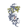
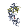
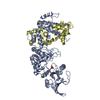
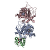
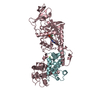
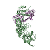
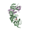
| Title | Crystal structure of the adenylyl cyclase domain of anthrax edema factor (EF) in complex with calmodulin and 2' deoxy, 3' anthraniloyl ATP | ||||||
 Components Components |
| ||||||
 Keywords Keywords | LYASE / helical domain / protein-protein complex | ||||||
| Function / homology |  Function and homology information Function and homology informationsymbiont-mediated perturbation of host signal transduction pathway / regulation of store-operated calcium channel activity / : / symbiont-mediated cAMP intoxication of host cell / DAPK1-calmodulin complex / : / calcium- and calmodulin-responsive adenylate cyclase activity / regulation of response to tumor cell / positive regulation of autophagic cell death / : ...symbiont-mediated perturbation of host signal transduction pathway / regulation of store-operated calcium channel activity / : / symbiont-mediated cAMP intoxication of host cell / DAPK1-calmodulin complex / : / calcium- and calmodulin-responsive adenylate cyclase activity / regulation of response to tumor cell / positive regulation of autophagic cell death / : / : / : / : / : / adenylate cyclase / : / type 3 metabotropic glutamate receptor binding / adenylate cyclase activity / establishment of protein localization to membrane / cAMP biosynthetic process / positive regulation of DNA binding / CaM pathway / Cam-PDE 1 activation / Sodium/Calcium exchangers / Calmodulin induced events / Reduction of cytosolic Ca++ levels / dATP binding / host cell cytosol / Activation of Ca-permeable Kainate Receptor / CREB1 phosphorylation through the activation of CaMKII/CaMKK/CaMKIV cascasde / Loss of phosphorylation of MECP2 at T308 / CREB1 phosphorylation through the activation of Adenylate Cyclase / negative regulation of high voltage-gated calcium channel activity / PKA activation / CaMK IV-mediated phosphorylation of CREB / Glycogen breakdown (glycogenolysis) / negative regulation of ryanodine-sensitive calcium-release channel activity / Activation of RAC1 downstream of NMDARs / organelle localization by membrane tethering / CLEC7A (Dectin-1) induces NFAT activation / : / autophagosome membrane docking / regulation of synaptic vesicle exocytosis / negative regulation of calcium ion export across plasma membrane / regulation of cardiac muscle cell action potential / presynaptic endocytosis / Synthesis of IP3 and IP4 in the cytosol / Phase 0 - rapid depolarisation / Negative regulation of NMDA receptor-mediated neuronal transmission / Unblocking of NMDA receptors, glutamate binding and activation / calcineurin-mediated signaling / RHO GTPases activate PAKs / nitric-oxide synthase binding / regulation of cell communication by electrical coupling involved in cardiac conduction / Ion transport by P-type ATPases / adenylate cyclase binding / Uptake and function of anthrax toxins / protein phosphatase activator activity / regulation of ryanodine-sensitive calcium-release channel activity / Long-term potentiation / Calcineurin activates NFAT / Regulation of MECP2 expression and activity / DARPP-32 events / Smooth Muscle Contraction / regulation of synaptic vesicle endocytosis / detection of calcium ion / catalytic complex / regulation of cardiac muscle contraction / cellular response to interferon-beta / RHO GTPases activate IQGAPs / positive regulation of nitric-oxide synthase activity / phosphatidylinositol 3-kinase binding / activation of adenylate cyclase activity / calcium channel inhibitor activity / presynaptic cytosol / Activation of AMPK downstream of NMDARs / regulation of release of sequestered calcium ion into cytosol by sarcoplasmic reticulum / enzyme regulator activity / eNOS activation / Ion homeostasis / Tetrahydrobiopterin (BH4) synthesis, recycling, salvage and regulation / regulation of calcium-mediated signaling / Protein methylation / titin binding / regulation of cardiac muscle contraction by regulation of the release of sequestered calcium ion / voltage-gated potassium channel complex / FCERI mediated Ca+2 mobilization / calcium channel complex / substantia nigra development / potassium ion transmembrane transport / regulation of heart rate / FCGR3A-mediated IL10 synthesis / calyx of Held / Ras activation upon Ca2+ influx through NMDA receptor / Antigen activates B Cell Receptor (BCR) leading to generation of second messengers / response to amphetamine / nitric-oxide synthase regulator activity / adenylate cyclase activator activity / protein serine/threonine kinase activator activity / VEGFR2 mediated cell proliferation Similarity search - Function | ||||||
| Biological species |   Homo sapiens (human) Homo sapiens (human) | ||||||
| Method |  X-RAY DIFFRACTION / SYNCHROTRON / FOURIER SYNTHESIS / Resolution: 3.6 Å X-RAY DIFFRACTION / SYNCHROTRON / FOURIER SYNTHESIS / Resolution: 3.6 Å | ||||||
 Authors Authors | Shen, Y. / Lee, Y.-S. / Soelaiman, S. / Bergson, P. / Lu, D. / Chen, A. / Beckingham, K. / Grabarek, Z. / Mrksich, M. / Tang, W.-J. | ||||||
 Citation Citation | Journal: Embo J. / Year: 2002 Title: Physiological calcium concentrations regulate calmodulin binding and catalysis of adenylyl cyclase exotoxins Authors: Shen, Y. / Lee, Y.-S. / Soelaiman, S. / Bergson, P. / Lu, D. / Chen, A. / Beckingham, K. / Grabarek, Z. / Mrksich, M. / Tang, W.-J. #1: Journal: Nature / Year: 2002Title: Structural basis for the activation of anthrax adenylyl cyclase exotoxin by calmodulin Authors: Drum, C.L. / Yan, S.Z. / Bard, J. / Shen, Y.Q. / Lu, D. / Soelaiman, S. / Grabarek, Z. / Bohm, A. / Tang, W.J. | ||||||
| History |
|
- Structure visualization
| Structure viewer | Molecule: MolmilJmol/JSmol |
|---|
- Downloads & links
-Download
| PDBx/mmCIF format | 1lvc.cif.gz | 389 KB | Display | PDBx/mmCIF format |
|---|---|---|---|---|
| PDB format | pdb1lvc.ent.gz | 313.2 KB | Display | PDB format |
| PDBx/mmJSON format | 1lvc.json.gz | Tree view | PDBx/mmJSON format | |
| Others |  Other downloads Other downloads |
-Validation report
| Arichive directory | https://data.pdbj.org/pub/pdb/validation_reports/lv/1lvcftp://data.pdbj.org/pub/pdb/validation_reports/lv/1lvc | HTTPS FTP |
|---|
-Related structure data
| Related structure data | 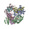 1k90S S: Starting model for refinement |
|---|---|
| Similar structure data |


-Links
 PDBj
PDBj


- Assembly
| Deposited unit | 
| ||||||||
|---|---|---|---|---|---|---|---|---|---|
| 1 | 
| ||||||||
| 2 | 
| ||||||||
| 3 | 
| ||||||||
| Unit cell |
| ||||||||
| Details | The biological assembly is monomer |
-Components
| #1: Protein | Mass: 58810.605 Da / Num. of mol.: 3 / Fragment: c-terminal domain (residues 291-800) Source method: isolated from a genetically manipulated source Source: (gene. exp.) #2: Protein | Mass: 16852.545 Da / Num. of mol.: 3 Source method: isolated from a genetically manipulated source Source: (gene. exp.) Homo sapiens (human) / Species (production host): Escherichia coli / Production host: #3: Chemical |   Mass: 173.040 Da / Num. of mol.: 3 / Source method: obtained synthetically / Formula: Yb Mass: 173.040 Da / Num. of mol.: 3 / Source method: obtained synthetically / Formula: Yb#4: Chemical |   Mass: 610.302 Da / Num. of mol.: 2 / Source method: obtained synthetically / Formula: C17H21N6O13P3 Mass: 610.302 Da / Num. of mol.: 2 / Source method: obtained synthetically / Formula: C17H21N6O13P3#5: Chemical | ChemComp-CA /   Mass: 40.078 Da / Num. of mol.: 6 / Source method: obtained synthetically / Formula: Ca Mass: 40.078 Da / Num. of mol.: 6 / Source method: obtained synthetically / Formula: Ca |
|---|
-Experimental details
-Experiment
| Experiment | Method: X-RAY DIFFRACTION / Number of used crystals: 1 |
|---|
- Sample preparation
| Crystal | Density Matthews: 3.69 Å3/Da / Density % sol: 66.71 % |
|---|---|
| Crystal grow | Temperature: 298 K / Method: vapor diffusion, hanging drop / pH: 6.5 Details: PEG 8000, ammonium sulfate, glycerol, pH 6.5, VAPOR DIFFUSION, HANGING DROP, temperature 298K |
| Crystal grow | *PLUS Method: unknownDetails: Hiratsuka, T., (1983) Biochem. Biophys. Acta., 742, 496. |
-Data collection
| Diffraction | Mean temperature: 200 K |
|---|---|
| Diffraction source | Source: SYNCHROTRON / Site: APS  / Beamline: 14-BM-C / Wavelength: 1 Å / Beamline: 14-BM-C / Wavelength: 1 Å |
| Detector | Type: ADSC QUANTUM 4 / Detector: CCD |
| Radiation | Monochromator: Graphite / Protocol: SINGLE WAVELENGTH / Monochromatic (M) / Laue (L): M / Scattering type: x-ray |
| Radiation wavelength | Wavelength: 1 Å / Relative weight: 1 |
| Reflection | Resolution: 3.5→29.96 Å / Num. all: 39156 / Num. obs: 38841 / % possible obs: 99.4 % / Observed criterion σ(F): 2 / Observed criterion σ(I): 2 / Redundancy: 7 % / Biso Wilson estimate: 46.7 Å2 / Rmerge(I) obs: 0.083 / Net I/σ(I): 26 |
| Reflection shell | Resolution: 3.6→3.73 Å / Redundancy: 7 % / Rmerge(I) obs: 0.356 / Mean I/σ(I) obs: 10 / Num. unique all: 3871 / % possible all: 99.9 |
| Reflection | *PLUS Highest resolution: 3.6 Å / Redundancy: 6.85 % |
- Processing
| Software |
| ||||||||||||||||||||||||||||||||||||
|---|---|---|---|---|---|---|---|---|---|---|---|---|---|---|---|---|---|---|---|---|---|---|---|---|---|---|---|---|---|---|---|---|---|---|---|---|---|
| Refinement | Method to determine structure: FOURIER SYNTHESIS Starting model: pdb entry 1K90 Resolution: 3.6→29.96 Å / Rfactor Rfree error: 0.007 / Data cutoff high absF: 457541.92 / Data cutoff high rms absF: 457541.92 / Isotropic thermal model: RESTRAINED / Cross valid method: THROUGHOUT / σ(F): 2 / Stereochemistry target values: Engh & Huber
| ||||||||||||||||||||||||||||||||||||
| Solvent computation | Solvent model: FLAT MODEL / Bsol: 51.5231 Å2 / ksol: 0.25238 e/Å3 | ||||||||||||||||||||||||||||||||||||
| Displacement parameters | Biso mean: 81.6 Å2
| ||||||||||||||||||||||||||||||||||||
| Refine analyze |
| ||||||||||||||||||||||||||||||||||||
| Refinement step | Cycle: LAST / Resolution: 3.6→29.96 Å
| ||||||||||||||||||||||||||||||||||||
| Refine LS restraints |
| ||||||||||||||||||||||||||||||||||||
| LS refinement shell | Resolution: 3.6→3.83 Å / Rfactor Rfree error: 0.019 / Total num. of bins used: 6
| ||||||||||||||||||||||||||||||||||||
| Xplor file |
| ||||||||||||||||||||||||||||||||||||
| Refinement | *PLUS Highest resolution: 3.6 Å / % reflection Rfree: 5 % | ||||||||||||||||||||||||||||||||||||
| Solvent computation | *PLUS | ||||||||||||||||||||||||||||||||||||
| Displacement parameters | *PLUS | ||||||||||||||||||||||||||||||||||||
| Refine LS restraints | *PLUS
|
