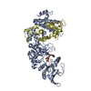
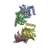
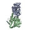
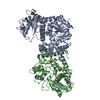
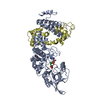
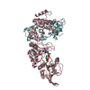
Entry Database : PDB / ID : 1sk6Title Crystal structure of the adenylyl cyclase domain of anthrax edema factor (EF) in complex with calmodulin, 3',5' cyclic AMP (cAMP), and pyrophosphate Calmodulin Calmodulin-sensitive adenylate cyclase Keywords / / / Function / homology Function Domain/homology Component
/ / / / / / / / / / / / / / / / / / / / / / / / / / / / / / / / / / / / / / / / / / / / / / / / / / / / / / / / / / / / / / / / / / / / / / / / / / / / / / / / / / / / / / / / / / / / / / / / / / / / / / / / / / / / / / / / / / / / / / / / / / / / / / / / / / / / / / / / / / / / / / / / / / / / / / / / Biological species Bacillus anthracis (anthrax bacterium)Homo sapiens (human)Method / / / Resolution : 3.2 Å Authors Guo, Q. / Shen, Y. / Zhukovskaya, N.L. / Tang, W.J. Journal : J.Biol.Chem. / Year : 2004Title : Structural and kinetic analyses of the interaction of anthrax adenylyl cyclase toxin with reaction products cAMP and pyrophosphate.Authors : Guo, Q. / Shen, Y. / Zhukovskaya, N.L. / Florian, J. / Tang, W.J. History Deposition Mar 4, 2004 Deposition site / Processing site Revision 1.0 Jun 8, 2004 Provider / Type Revision 1.1 Apr 29, 2008 Group Revision 1.2 Jul 13, 2011 Group Revision 1.3 Nov 20, 2019 Group / Derived calculationsCategory database_PDB_caveat / pdbx_struct_conn_angle ... database_PDB_caveat / pdbx_struct_conn_angle / pdbx_validate_close_contact / struct_conn Revision 1.4 Aug 23, 2023 Group Data collection / Database references ... Data collection / Database references / Derived calculations / Refinement description Category chem_comp_atom / chem_comp_bond ... chem_comp_atom / chem_comp_bond / database_2 / pdbx_initial_refinement_model / pdbx_struct_conn_angle / struct_conn / struct_site Item _database_2.pdbx_DOI / _database_2.pdbx_database_accession ... _database_2.pdbx_DOI / _database_2.pdbx_database_accession / _pdbx_struct_conn_angle.ptnr1_auth_comp_id / _pdbx_struct_conn_angle.ptnr1_auth_seq_id / _pdbx_struct_conn_angle.ptnr1_label_asym_id / _pdbx_struct_conn_angle.ptnr1_label_atom_id / _pdbx_struct_conn_angle.ptnr1_label_comp_id / _pdbx_struct_conn_angle.ptnr1_label_seq_id / _pdbx_struct_conn_angle.ptnr2_auth_seq_id / _pdbx_struct_conn_angle.ptnr2_label_asym_id / _pdbx_struct_conn_angle.ptnr3_auth_comp_id / _pdbx_struct_conn_angle.ptnr3_auth_seq_id / _pdbx_struct_conn_angle.ptnr3_label_asym_id / _pdbx_struct_conn_angle.ptnr3_label_atom_id / _pdbx_struct_conn_angle.ptnr3_label_comp_id / _pdbx_struct_conn_angle.ptnr3_label_seq_id / _pdbx_struct_conn_angle.value / _struct_conn.pdbx_dist_value / _struct_conn.ptnr1_auth_asym_id / _struct_conn.ptnr1_auth_comp_id / _struct_conn.ptnr1_auth_seq_id / _struct_conn.ptnr1_label_asym_id / _struct_conn.ptnr1_label_atom_id / _struct_conn.ptnr1_label_comp_id / _struct_conn.ptnr1_label_seq_id / _struct_conn.ptnr2_auth_asym_id / _struct_conn.ptnr2_auth_comp_id / _struct_conn.ptnr2_auth_seq_id / _struct_conn.ptnr2_label_asym_id / _struct_conn.ptnr2_label_atom_id / _struct_conn.ptnr2_label_comp_id / _struct_site.pdbx_auth_asym_id / _struct_site.pdbx_auth_comp_id / _struct_site.pdbx_auth_seq_id
Show all Show less Remark 999 SEQUENCE Residue (B ASN 521 ) and Residue (B GLU 524 ) are linked together. Residue (C PHE 773 ) ... SEQUENCE Residue (B ASN 521 ) and Residue (B GLU 524 ) are linked together. Residue (C PHE 773 ) and Residue (C LYS 774 ) are not linked, distance of C-N bond is 1.80.
 Movie
Movie Controller

 Yorodumi
 Open data
 Basic information
 Components
 Keywords
 Function and homology information
 Homo sapiens (human)
 X-RAY DIFFRACTION /
  Authors
 Citation
 Structure visualization
 Downloads & links
 Other downloads
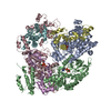


 PDBj
PDBj


 Assembly


 Mass: 173.040 Da / Num. of mol.: 9 / Source method: obtained synthetically / Formula: Yb
 Mass: 329.206 Da / Num. of mol.: 3 / Source method: obtained synthetically / Formula: C10H12N5O6P
 Mass: 175.959 Da / Num. of mol.: 3 / Source method: obtained synthetically / Formula: H2O7P2
 Mass: 40.078 Da / Num. of mol.: 6 / Source method: obtained synthetically / Formula: Ca
 Sample preparation
 / Beamline: 19-ID / Wavelength: 1 Å
 Processing