- PDB-2yjn: Structure of the glycosyltransferase EryCIII from the erythromyci... -
+
Open data
ID or keywords:
Loading...
-
Basic information
Entry
Database: PDB / ID: 2yjn
Title
Structure of the glycosyltransferase EryCIII from the erythromycin biosynthetic pathway, in complex with its activating partner, EryCII
Components
DTDP-4-KETO-6-DEOXY-HEXOSE 3,4-ISOMERASE
GLYCOSYLTRANSFERASE
Keywords
TRANSFERASE / CYTOCHROME P450
Function / homology
Function and homology information
3-alpha-mycarosylerythronolide B desosaminyl transferase / UDP-glycosyltransferase activity / hexosyltransferase activity / oxidoreductase activity, acting on paired donors, with incorporation or reduction of molecular oxygen / antibiotic biosynthetic process / monooxygenase activity / iron ion binding / heme binding Similarity search - Function
Cytochrome P450 family protein EryCII / 3-alpha-mycarosylerythronolide B desosaminyl transferase / 3-alpha-mycarosylerythronolide B desosaminyl transferase / Cytochrome P450 family protein EryCII Similarity search - Component
Biological species
SACCHAROPOLYSPORA ERYTHRAEA (bacteria)
Method
X-RAY DIFFRACTION / SYNCHROTRON / SAD / Resolution: 3.091 Å
Protocol: SINGLE WAVELENGTH / Monochromatic (M) / Laue (L): M / Scattering type: x-ray
Radiation wavelength
Wavelength: 0.9756 Å / Relative weight: 1
Reflection
Resolution: 3.1→20 Å / Num. obs: 17457 / % possible obs: 99.4 % / Observed criterion σ(I): 2 / Redundancy: 7.2 % / Biso Wilson estimate: 93.73 Å2 / Rmerge(I) obs: 0.04 / Net I/σ(I): 14
Reflection shell
Resolution: 3.1→3.1 Å / Redundancy: 7.5 % / Rmerge(I) obs: 0.27 / Mean I/σ(I) obs: 3 / % possible all: 100
-
Processing
Software
Name
Version
Classification
PHENIX
(PHENIX.REFINE)
refinement
XDS
datareduction
SCALA
datascaling
autoSHARP
phasing
Refinement
Method to determine structure: SAD Starting model: NONE Resolution: 3.091→19.677 Å / SU ML: 0.38 / σ(F): 1.33 / Phase error: 27.99 / Stereochemistry target values: ML Details: CHAIN A RESIDUES 1-18 ARE MISSING CHAIN B RESIDUES 1-19 ARE MISSING
Rfactor
Num. reflection
% reflection
Rfree
0.2615
891
5.1 %
Rwork
0.2102
-
-
obs
0.2129
17457
98.84 %
Solvent computation
Shrinkage radii: 0.27 Å / VDW probe radii: 0.6 Å / Solvent model: FLAT BULK SOLVENT MODEL / Bsol: 74.51 Å2 / ksol: 0.321 e/Å3
 Movie
Movie Controller

 Yorodumi
 Open data
 Basic information
 Components
 Keywords
 Function and homology information
 SACCHAROPOLYSPORA ERYTHRAEA (bacteria)
 X-RAY DIFFRACTION /
  Authors
 Citation
 Structure visualization
 Downloads & links
 Other downloads
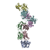


 PDBj
PDBj
 Assembly
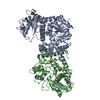

 Sample preparation
 / Beamline: ID29 / Wavelength: 0.9756
  Processing