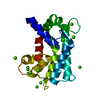
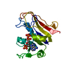
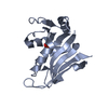
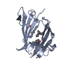
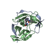
[English] 日本語
Yorodumi
- PDB-1ctr: DRUG BINDING BY CALMODULIN: CRYSTAL STRUCTURE OF A CALMODULIN-TRI... -

+
Open data


ID or keywords:

Loading...

-
Basic information

Entry
Database: PDB / ID: 1ctr
TitleDRUG BINDING BY CALMODULIN: CRYSTAL STRUCTURE OF A CALMODULIN-TRIFLUOPERAZINE COMPLEX
ComponentsCALMODULIN
KeywordsCALCIUM-BINDING PROTEIN
Function / homology
Function and homology information


: / : / positive regulation of cyclic-nucleotide phosphodiesterase activity / : / establishment of protein localization to mitochondrial membrane / type 3 metabotropic glutamate receptor binding / CaM pathway / Cam-PDE 1 activation / Sodium/Calcium exchangers / Calmodulin induced events ...: / : / positive regulation of cyclic-nucleotide phosphodiesterase activity / : / establishment of protein localization to mitochondrial membrane / type 3 metabotropic glutamate receptor binding / CaM pathway / Cam-PDE 1 activation / Sodium/Calcium exchangers / Calmodulin induced events / Reduction of cytosolic Ca++ levels / Activation of Ca-permeable Kainate Receptor / CREB1 phosphorylation through the activation of CaMKII/CaMKK/CaMKIV cascasde / Loss of phosphorylation of MECP2 at T308 / CREB1 phosphorylation through the activation of Adenylate Cyclase / PKA activation / negative regulation of high voltage-gated calcium channel activity / CaMK IV-mediated phosphorylation of CREB / Glycogen breakdown (glycogenolysis) / nitric-oxide synthase binding / response to corticosterone / positive regulation of DNA binding / organelle localization by membrane tethering / negative regulation of calcium ion export across plasma membrane / CLEC7A (Dectin-1) induces NFAT activation / Activation of RAC1 downstream of NMDARs / regulation of synaptic vesicle exocytosis / mitochondrion-endoplasmic reticulum membrane tethering / autophagosome membrane docking / presynaptic endocytosis / regulation of cardiac muscle cell action potential / negative regulation of peptidyl-threonine phosphorylation / calcineurin-mediated signaling / positive regulation of ryanodine-sensitive calcium-release channel activity / regulation of cell communication by electrical coupling involved in cardiac conduction / Synthesis of IP3 and IP4 in the cytosol / negative regulation of ryanodine-sensitive calcium-release channel activity / Phase 0 - rapid depolarisation / Negative regulation of NMDA receptor-mediated neuronal transmission / Unblocking of NMDA receptors, glutamate binding and activation / regulation of synaptic vesicle endocytosis / RHO GTPases activate PAKs / Ion transport by P-type ATPases / Uptake and function of anthrax toxins / protein phosphatase activator activity / adenylate cyclase binding / Long-term potentiation / Regulation of MECP2 expression and activity / Calcineurin activates NFAT / regulation of ryanodine-sensitive calcium-release channel activity / DARPP-32 events / catalytic complex / Smooth Muscle Contraction / regulation of cardiac muscle contraction / detection of calcium ion / RHO GTPases activate IQGAPs / phosphatidylinositol 3-kinase binding / calcium channel inhibitor activity / regulation of cardiac muscle contraction by regulation of the release of sequestered calcium ion / presynaptic cytosol / cellular response to interferon-beta / Protein methylation / Activation of AMPK downstream of NMDARs / activation of adenylate cyclase activity / positive regulation of protein autophosphorylation / regulation of release of sequestered calcium ion into cytosol by sarcoplasmic reticulum / Ion homeostasis / eNOS activation / enzyme regulator activity / regulation of calcium-mediated signaling / titin binding / Tetrahydrobiopterin (BH4) synthesis, recycling, salvage and regulation / voltage-gated potassium channel complex / sperm midpiece / calcium channel complex / substantia nigra development / positive regulation of peptidyl-threonine phosphorylation / calyx of Held / nitric-oxide synthase regulator activity / response to amphetamine / FCERI mediated Ca+2 mobilization / adenylate cyclase activator activity / Ras activation upon Ca2+ influx through NMDA receptor / FCGR3A-mediated IL10 synthesis / regulation of heart rate / sarcomere / positive regulation of nitric-oxide synthase activity / protein serine/threonine kinase activator activity / Antigen activates B Cell Receptor (BCR) leading to generation of second messengers / regulation of cytokinesis / VEGFR2 mediated cell proliferation / VEGFR2 mediated vascular permeability / Translocation of SLC2A4 (GLUT4) to the plasma membrane / positive regulation of protein serine/threonine kinase activity / spindle microtubule / positive regulation of receptor signaling pathway via JAK-STAT / RAF activation / Transcriptional activation of mitochondrial biogenesis / mitochondrial membrane / Stimuli-sensing channels
Similarity search - Function
: / EF-hand / Recoverin; domain 1 / EF-hand domain pair / EF-hand, calcium binding motif / EF-Hand 1, calcium-binding site / EF-hand calcium-binding domain. / EF-hand calcium-binding domain profile. / EF-hand domain / EF-hand domain pair ...: / EF-hand / Recoverin; domain 1 / EF-hand domain pair / EF-hand, calcium binding motif / EF-Hand 1, calcium-binding site / EF-hand calcium-binding domain. / EF-hand calcium-binding domain profile. / EF-hand domain / EF-hand domain pair / Orthogonal Bundle / Mainly Alpha
Similarity search - Domain/homology
Chem-TFP / Calmodulin-1 / Calmodulin-3
Similarity search - Component
Biological speciesHomo sapiens (human)
MethodX-RAY DIFFRACTION / Resolution: 2.45 Å
AuthorsCook, W.J. / Walter, L.J. / Walter, M.R.
CitationJournal: Biochemistry / Year: 1994
Title: Drug binding by calmodulin: crystal structure of a calmodulin-trifluoperazine complex.
Authors: Cook, W.J. / Walter, L.J. / Walter, M.R.
History
DepositionSep 21, 1994Processing site: BNL
Revision 1.0Dec 20, 1994Provider: repository / Type: Initial release
Revision 1.1Mar 24, 2008Group: Version format compliance
Revision 1.2Jul 13, 2011Group: Version format compliance
Revision 1.3Feb 7, 2024Group: Data collection / Database references ...Data collection / Database references / Derived calculations / Other
Category: chem_comp_atom / chem_comp_bond ...chem_comp_atom / chem_comp_bond / database_2 / pdbx_database_status / pdbx_struct_conn_angle / struct_conn / struct_site
Item: _database_2.pdbx_DOI / _database_2.pdbx_database_accession ..._database_2.pdbx_DOI / _database_2.pdbx_database_accession / _pdbx_database_status.process_site / _pdbx_struct_conn_angle.ptnr1_auth_comp_id / _pdbx_struct_conn_angle.ptnr1_auth_seq_id / _pdbx_struct_conn_angle.ptnr1_label_atom_id / _pdbx_struct_conn_angle.ptnr1_label_comp_id / _pdbx_struct_conn_angle.ptnr1_label_seq_id / _pdbx_struct_conn_angle.ptnr3_auth_comp_id / _pdbx_struct_conn_angle.ptnr3_auth_seq_id / _pdbx_struct_conn_angle.ptnr3_label_atom_id / _pdbx_struct_conn_angle.ptnr3_label_comp_id / _pdbx_struct_conn_angle.ptnr3_label_seq_id / _pdbx_struct_conn_angle.value / _struct_conn.pdbx_dist_value / _struct_conn.ptnr1_auth_comp_id / _struct_conn.ptnr1_auth_seq_id / _struct_conn.ptnr1_label_asym_id / _struct_conn.ptnr1_label_atom_id / _struct_conn.ptnr1_label_comp_id / _struct_conn.ptnr1_label_seq_id / _struct_conn.ptnr2_auth_comp_id / _struct_conn.ptnr2_auth_seq_id / _struct_conn.ptnr2_label_asym_id / _struct_conn.ptnr2_label_atom_id / _struct_conn.ptnr2_label_comp_id / _struct_conn.ptnr2_label_seq_id / _struct_site.pdbx_auth_asym_id / _struct_site.pdbx_auth_comp_id / _struct_site.pdbx_auth_seq_id

-
Structure visualization

Structure viewerMolecule:
MolmilJmol/JSmol

Downloads & links

-
Assembly

Deposited unit
A: CALMODULIN
hetero molecules


Theoretical massNumber of molelcules
Total (without water)17,2896
Polymers16,7211
Non-polymers5685
Water00
1


  • Idetical with deposited unit
  • defined by author
TypeNameSymmetry operationNumber
identity operation1_555x,y,z1
Unit cell
Length a, b, c (Å)41.000, 41.000, 178.900
Angle α, β, γ (deg.)90.00, 90.00, 120.00
Int Tables number154
Space group name H-MP3221

-
Components

#1: Protein CALMODULIN


Mass: 16721.350 Da / Num. of mol.: 1
Source method: isolated from a genetically manipulated source
Source: (gene. exp.) Homo sapiens (human) / References: UniProt: P62158, UniProt: P0DP23*PLUS
#2: Chemical
ChemComp-CA / CALCIUM ION


Mass: 40.078 Da / Num. of mol.: 4 / Source method: obtained synthetically / Formula: Ca
#3: Chemical ChemComp-TFP / 10-[3-(4-METHYL-PIPERAZIN-1-YL)-PROPYL]-2-TRIFLUOROMETHYL-10H-PHENOTHIAZINE


Mass: 407.496 Da / Num. of mol.: 1 / Source method: obtained synthetically / Formula: C21H24F3N3S / Comment: antipsychotic*YM

-
Experimental details

-
Experiment

ExperimentMethod: X-RAY DIFFRACTION

-
Sample preparation

CrystalDensity Matthews: 2.6 Å3/Da / Density % sol: 52.61 %
Crystal grow
*PLUS
Temperature: 4 ℃ / pH: 5.8 / Method: vapor diffusion, hanging drop
Components of the solutions
*PLUS
IDConc.Common nameCrystal-IDSol-ID
220 %PEG-MME 20001drop
30.1 Msodium cacodylate1drop
420 %PEG-MME 20001reservoir
50.1 Msodium cacodylate1reservoir
1CaM-TFP1drop

-
Data collection

RadiationScattering type: x-ray
Radiation wavelengthRelative weight: 1
Reflection
*PLUS
Highest resolution: 2.45 Å / Num. obs: 6240 / % possible obs: 89 % / Num. measured all: 16284 / Rmerge(I) obs: 0.092

-
Processing

Software
NameClassification
X-PLORmodel building
X-PLORrefinement
X-PLORphasing
RefinementResolution: 2.45→6 Å / σ(F): 2 /
RfactorNum. reflection
Rwork0.222 -
obs0.222 5060
Refinement stepCycle: LAST / Resolution: 2.45→6 Å
ProteinNucleic acidLigandSolventTotal
Num. atoms1109 0 32 0 1141
Refine LS restraints
Refine-IDTypeDev ideal
X-RAY DIFFRACTIONx_bond_d0.015
X-RAY DIFFRACTIONx_bond_d_na
X-RAY DIFFRACTIONx_bond_d_prot
X-RAY DIFFRACTIONx_angle_d
X-RAY DIFFRACTIONx_angle_d_na
X-RAY DIFFRACTIONx_angle_d_prot
X-RAY DIFFRACTIONx_angle_deg2.5
X-RAY DIFFRACTIONx_angle_deg_na
X-RAY DIFFRACTIONx_angle_deg_prot
X-RAY DIFFRACTIONx_dihedral_angle_d
X-RAY DIFFRACTIONx_dihedral_angle_d_na
X-RAY DIFFRACTIONx_dihedral_angle_d_prot
X-RAY DIFFRACTIONx_improper_angle_d
X-RAY DIFFRACTIONx_improper_angle_d_na
X-RAY DIFFRACTIONx_improper_angle_d_prot
X-RAY DIFFRACTIONx_mcbond_it
X-RAY DIFFRACTIONx_mcangle_it
X-RAY DIFFRACTIONx_scbond_it
X-RAY DIFFRACTIONx_scangle_it

+
About Yorodumi

-
News

-
Feb 9, 2022. New format data for meta-information of EMDB entries

New format data for meta-information of EMDB entries

  • Version 3 of the EMDB header file is now the official format.
  • The previous official version 1.9 will be removed from the archive.

Related info.:EMDB header

External links:wwPDB to switch to version 3 of the EMDB data model

-
Aug 12, 2020. Covid-19 info

Covid-19 info

URL: https://pdbj.org/emnavi/covid19.php

New page: Covid-19 featured information page in EM Navigator.

Related info.:Covid-19 info / Mar 5, 2020. Novel coronavirus structure data

+
Mar 5, 2020. Novel coronavirus structure data

Novel coronavirus structure data

Related info.:Yorodumi Speices / Aug 12, 2020. Covid-19 info

External links:COVID-19 featured content - PDBj / Molecule of the Month (242):Coronavirus Proteases

+
Jan 31, 2019. EMDB accession codes are about to change! (news from PDBe EMDB page)

EMDB accession codes are about to change! (news from PDBe EMDB page)

  • The allocation of 4 digits for EMDB accession codes will soon come to an end. Whilst these codes will remain in use, new EMDB accession codes will include an additional digit and will expand incrementally as the available range of codes is exhausted. The current 4-digit format prefixed with “EMD-” (i.e. EMD-XXXX) will advance to a 5-digit format (i.e. EMD-XXXXX), and so on. It is currently estimated that the 4-digit codes will be depleted around Spring 2019, at which point the 5-digit format will come into force.
  • The EM Navigator/Yorodumi systems omit the EMD- prefix.

Related info.:Q: What is EMD? / ID/Accession-code notation in Yorodumi/EM Navigator

External links:EMDB Accession Codes are Changing Soon! / Contact to PDBj

+
Jul 12, 2017. Major update of PDB

Major update of PDB

  • wwPDB released updated PDB data conforming to the new PDBx/mmCIF dictionary.
  • This is a major update changing the version number from 4 to 5, and with Remediation, in which all the entries are updated.
  • In this update, many items about electron microscopy experimental information are reorganized (e.g. em_software).
  • Now, EM Navigator and Yorodumi are based on the updated data.

External links:wwPDB Remediation / Enriched Model Files Conforming to OneDep Data Standards Now Available in the PDB FTP Archive

-
Yorodumi

Thousand views of thousand structures

  • Yorodumi is a browser for structure data from EMDB, PDB, SASBDB, etc.
  • This page is also the successor to EM Navigator detail page, and also detail information page/front-end page for Omokage search.
  • The word "yorodu" (or yorozu) is an old Japanese word meaning "ten thousand". "mi" (miru) is to see.

Related info.:EMDB / PDB / SASBDB / Comparison of 3 databanks / Yorodumi Search / Aug 31, 2016. New EM Navigator & Yorodumi / Yorodumi Papers / Jmol/JSmol / Function and homology information / Changes in new EM Navigator and Yorodumi

Read more