 Movie
Movie Controller

[English] 日本語
 Yorodumi
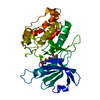
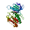
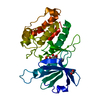
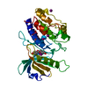
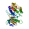
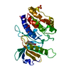
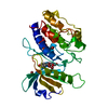
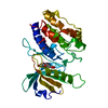
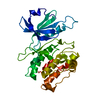
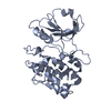
- PDB-1jkk: 2.4A X-RAY STRUCTURE OF TERNARY COMPLEX OF A CATALYTIC DOMAIN OF ... -
+ Open data

- Basic information
| Entry | Database: PDB / ID: 1jkk | ||||||
|---|---|---|---|---|---|---|---|
| Title | 2.4A X-RAY STRUCTURE OF TERNARY COMPLEX OF A CATALYTIC DOMAIN OF DEATH-ASSOCIATED PROTEIN KINASE WITH ATP ANALOGUE AND MG. | ||||||
 Components Components | DEATH-ASSOCIATED PROTEIN KINASE | ||||||
 Keywords Keywords | TRANSFERASE | ||||||
| Function / homology |  Function and homology information Function and homology informationcellular response to hydroperoxide / negative regulation of kinase activity / regulation of response to tumor cell / positive regulation of autophagic cell death / Caspase activation via Dependence Receptors in the absence of ligand / defense response to tumor cell / calcium/calmodulin-dependent protein kinase activity / regulation of NMDA receptor activity / syntaxin-1 binding / positive regulation of autophagy ...cellular response to hydroperoxide / negative regulation of kinase activity / regulation of response to tumor cell / positive regulation of autophagic cell death / Caspase activation via Dependence Receptors in the absence of ligand / defense response to tumor cell / calcium/calmodulin-dependent protein kinase activity / regulation of NMDA receptor activity / syntaxin-1 binding / positive regulation of autophagy / regulation of autophagy / apoptotic signaling pathway / cellular response to type II interferon / protein autophosphorylation / actin cytoskeleton / regulation of apoptotic process / protein phosphorylation / protein kinase activity / calmodulin binding / non-specific serine/threonine protein kinase / negative regulation of translation / postsynaptic density / intracellular signal transduction / positive regulation of apoptotic process / protein serine kinase activity / protein serine/threonine kinase activity / apoptotic process / negative regulation of apoptotic process / GTP binding / glutamatergic synapse / ATP binding / identical protein binding / nucleus / plasma membrane / cytoplasm Similarity search - Function | ||||||
| Biological species |  Homo sapiens (human) Homo sapiens (human) | ||||||
| Method |  X-RAY DIFFRACTION / SYNCHROTRON / MOLECULAR REPLACEMENT / Resolution: 2.4 Å X-RAY DIFFRACTION / SYNCHROTRON / MOLECULAR REPLACEMENT / Resolution: 2.4 Å | ||||||
 Authors Authors | Tereshko, V. / Teplova, M. / Brunzelle, J. / Watterson, D.M. / Egli, M. | ||||||
 Citation Citation | Journal: Nat.Struct.Biol. / Year: 2001 Title: Crystal structures of the catalytic domain of human protein kinase associated with apoptosis and tumor suppression. Authors: Tereshko, V. / Teplova, M. / Brunzelle, J. / Watterson, D.M. / Egli, M. | ||||||
| History |
|
- Structure visualization
| Structure viewer | Molecule: MolmilJmol/JSmol |
|---|
- Downloads & links
-Download
| PDBx/mmCIF format | 1jkk.cif.gz | 72.2 KB | Display | PDBx/mmCIF format |
|---|---|---|---|---|
| PDB format | pdb1jkk.ent.gz | 52.3 KB | Display | PDB format |
| PDBx/mmJSON format | 1jkk.json.gz | Tree view | PDBx/mmJSON format | |
| Others |  Other downloads Other downloads |
-Validation report
| Arichive directory | https://data.pdbj.org/pub/pdb/validation_reports/jk/1jkkftp://data.pdbj.org/pub/pdb/validation_reports/jk/1jkk | HTTPS FTP |
|---|
-Related structure data


-Links
 PDBj
PDBj


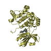
- Assembly
| Deposited unit | 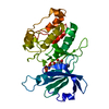
| ||||||||
|---|---|---|---|---|---|---|---|---|---|
| 1 |
| ||||||||
| Unit cell |
|
-Components
| #1: Protein | Mass: 33794.367 Da / Num. of mol.: 1 / Fragment: CATALYTIC DOMAIN, PROTEIN KINASE DOMAIN Source method: isolated from a genetically manipulated source Source: (gene. exp.) Homo sapiens (human) / Production host:  References: UniProt: P53355, Transferases; Transferring phosphorus-containing groups; Phosphotransferases with an alcohol group as acceptor |
|---|---|
| #2: Chemical | ChemComp-MG /   Mass: 24.305 Da / Num. of mol.: 1 / Source method: obtained synthetically / Formula: Mg Mass: 24.305 Da / Num. of mol.: 1 / Source method: obtained synthetically / Formula: Mg |
| #3: Chemical | ChemComp-ANP /   Mass: 506.196 Da / Num. of mol.: 1 / Source method: obtained synthetically / Formula: C10H17N6O12P3 / Comment: AMP-PNP, energy-carrying molecule analogue*YM Mass: 506.196 Da / Num. of mol.: 1 / Source method: obtained synthetically / Formula: C10H17N6O12P3 / Comment: AMP-PNP, energy-carrying molecule analogue*YM |
| #4: Water | ChemComp-HOH /  Mass: 18.015 Da / Num. of mol.: 127 / Source method: isolated from a natural source / Formula: H2O Mass: 18.015 Da / Num. of mol.: 127 / Source method: isolated from a natural source / Formula: H2O |
-Experimental details
-Experiment
| Experiment | Method: X-RAY DIFFRACTION / Number of used crystals: 1 |
|---|
- Sample preparation
| Crystal | Density Matthews: 1.91 Å3/Da / Density % sol: 35.56 % | |||||||||||||||||||||||||||||||||||||||||||||||||||||||||||||||
|---|---|---|---|---|---|---|---|---|---|---|---|---|---|---|---|---|---|---|---|---|---|---|---|---|---|---|---|---|---|---|---|---|---|---|---|---|---|---|---|---|---|---|---|---|---|---|---|---|---|---|---|---|---|---|---|---|---|---|---|---|---|---|---|---|
| Crystal grow | pH: 7.5 Details: PROTEIN, AMPPnP, AMMONIUM SULFATE, TRIS-HL (pH 8), MGCL2 SOAKING, pH 7.5 | |||||||||||||||||||||||||||||||||||||||||||||||||||||||||||||||
| Crystal grow | *PLUS Method: vapor diffusion, hanging drop | |||||||||||||||||||||||||||||||||||||||||||||||||||||||||||||||
| Components of the solutions | *PLUS
|
-Data collection
| Diffraction | Mean temperature: 100 K |
|---|---|
| Diffraction source | Source: SYNCHROTRON / Site: APS  / Beamline: 17-ID / Wavelength: 0.984 / Beamline: 17-ID / Wavelength: 0.984 |
| Detector | Type: MARRESEARCH / Detector: CCD / Date: Sep 21, 2000 |
| Radiation | Protocol: SINGLE WAVELENGTH / Monochromatic (M) / Laue (L): M / Scattering type: x-ray |
| Radiation wavelength | Wavelength: 0.984 Å / Relative weight: 1 |
| Reflection | Resolution: 2.4→20 Å / Num. obs: 10555 / % possible obs: 99.8 % / Observed criterion σ(I): -3 / Redundancy: 4.5 % / Biso Wilson estimate: 25.6 Å2 / Rmerge(I) obs: 0.07 |
| Reflection shell | Resolution: 2.4→2.49 Å / Rmerge(I) obs: 0.3 / % possible all: 99.9 |
| Reflection | *PLUS Highest resolution: 2.4 Å / Lowest resolution: 20 Å / Rmerge(I) obs: 0.07 |
| Reflection shell | *PLUS % possible obs: 99.9 % / Num. unique obs: 1033 / Rmerge(I) obs: 0.3 |
- Processing
| Software |
| ||||||||||||||||||||||||||||||||||||||||||||||||||||||||||||||||||||||||||||||||
|---|---|---|---|---|---|---|---|---|---|---|---|---|---|---|---|---|---|---|---|---|---|---|---|---|---|---|---|---|---|---|---|---|---|---|---|---|---|---|---|---|---|---|---|---|---|---|---|---|---|---|---|---|---|---|---|---|---|---|---|---|---|---|---|---|---|---|---|---|---|---|---|---|---|---|---|---|---|---|---|---|---|
| Refinement | Method to determine structure: MOLECULAR REPLACEMENT / Resolution: 2.4→19.59 Å / Rfactor Rfree error: 0.008 / Data cutoff high absF: 624571.44 / Data cutoff low absF: 0 / Isotropic thermal model: RESTRAINED / Cross valid method: THROUGHOUT / σ(F): 0
| ||||||||||||||||||||||||||||||||||||||||||||||||||||||||||||||||||||||||||||||||
| Solvent computation | Solvent model: FLAT MODEL / Bsol: 41.8519 Å2 / ksol: 0.39953 e/Å3 | ||||||||||||||||||||||||||||||||||||||||||||||||||||||||||||||||||||||||||||||||
| Displacement parameters | Biso mean: 28.5 Å2
| ||||||||||||||||||||||||||||||||||||||||||||||||||||||||||||||||||||||||||||||||
| Refine analyze |
| ||||||||||||||||||||||||||||||||||||||||||||||||||||||||||||||||||||||||||||||||
| Refinement step | Cycle: LAST / Resolution: 2.4→19.59 Å
| ||||||||||||||||||||||||||||||||||||||||||||||||||||||||||||||||||||||||||||||||
| Refine LS restraints |
| ||||||||||||||||||||||||||||||||||||||||||||||||||||||||||||||||||||||||||||||||
| LS refinement shell | Resolution: 2.4→2.55 Å / Rfactor Rfree error: 0.022 / Total num. of bins used: 6
| ||||||||||||||||||||||||||||||||||||||||||||||||||||||||||||||||||||||||||||||||
| Refinement | *PLUS Num. reflection obs: 9888 / Num. reflection Rfree: 1011 / Rfactor obs: 0.201 / Rfactor Rfree: 0.239 / Rfactor Rwork: 0.201 | ||||||||||||||||||||||||||||||||||||||||||||||||||||||||||||||||||||||||||||||||
| Solvent computation | *PLUS | ||||||||||||||||||||||||||||||||||||||||||||||||||||||||||||||||||||||||||||||||
| Displacement parameters | *PLUS | ||||||||||||||||||||||||||||||||||||||||||||||||||||||||||||||||||||||||||||||||
| Refine LS restraints | *PLUS
| ||||||||||||||||||||||||||||||||||||||||||||||||||||||||||||||||||||||||||||||||
| LS refinement shell | *PLUS Rfactor Rfree: 0.266 / Rfactor Rwork: 0.234 |
