Resolution: 2.4→30 Å / Num. obs: 10558 / % possible obs: 98.6 % / Redundancy: 8.8 % / Biso Wilson estimate: 47.9 Å2 / Rmerge(I) obs: 0.064 / Net I/σ(I): 18.9
Reflection shell
Resolution: 2.4→2.49 Å / Rmerge(I) obs: 0.38 / Mean I/σ(I) obs: 3.5 / % possible all: 98.2
-
Processing
Software
Name
Version
Classification
CNS
1.1
refinement
DENZO
datareduction
SCALEPACK
datascaling
SHARP
phasing
Refinement
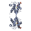
Method to determine structure: MAD / Resolution: 2.4→19.88 Å / Rfactor Rfree error: 0.011 / Data cutoff high absF: 1351678.82 / Data cutoff low absF: 0 / Isotropic thermal model: RESTRAINED / Cross valid method: THROUGHOUT / σ(F): 2 Details: The N-terminal domain is locked in a closed conformation with a disulfide bond. This prevents the glutamate in the 12th position of the loop from interacting with the calcium ion in sites I ...Details: The N-terminal domain is locked in a closed conformation with a disulfide bond. This prevents the glutamate in the 12th position of the loop from interacting with the calcium ion in sites I and II. Bond angles in residues 60, 61, 94 and 132 deviate by more than 6*RMSD relative to the standard dictionary (see REMARK 500). These residues are located in the calcium binding loops, thus their conformation is constrained by the calcium ions.
In the structure databanks used in Yorodumi, some data are registered as the other names, "COVID-19 virus" and "2019-nCoV". Here are the details of the virus and the list of structure data.
Jan 31, 2019. EMDB accession codes are about to change! (news from PDBe EMDB page)
EMDB accession codes are about to change! (news from PDBe EMDB page)
The allocation of 4 digits for EMDB accession codes will soon come to an end. Whilst these codes will remain in use, new EMDB accession codes will include an additional digit and will expand incrementally as the available range of codes is exhausted. The current 4-digit format prefixed with “EMD-” (i.e. EMD-XXXX) will advance to a 5-digit format (i.e. EMD-XXXXX), and so on. It is currently estimated that the 4-digit codes will be depleted around Spring 2019, at which point the 5-digit format will come into force.
The EM Navigator/Yorodumi systems omit the EMD- prefix.
Related info.:Q: What is EMD? / ID/Accession-code notation in Yorodumi/EM Navigator
Yorodumi is a browser for structure data from EMDB, PDB, SASBDB, etc.
This page is also the successor to EM Navigator detail page, and also detail information page/front-end page for Omokage search.
The word "yorodu" (or yorozu) is an old Japanese word meaning "ten thousand". "mi" (miru) is to see.
Related info.:EMDB / PDB / SASBDB / Comparison of 3 databanks / Yorodumi Search / Aug 31, 2016. New EM Navigator & Yorodumi / Yorodumi Papers / Jmol/JSmol / Function and homology information / Changes in new EM Navigator and Yorodumi
 Movie
Movie Controller


 Open data
 Basic information
 Components
 Keywords
 Function and homology information
 Homo sapiens (human)
 X-RAY DIFFRACTION /
  Authors
 Citation
 Structure visualization
 Downloads & links
 Other downloads
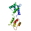


 PDBj
PDBj


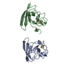
 Assembly


 Mass: 40.078 Da / Num. of mol.: 4 / Source method: obtained synthetically / Formula: Ca
 Mass: 118.174 Da / Num. of mol.: 1 / Source method: obtained synthetically / Formula: C6H14O2 / Comment: precipitant*YM
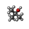
 Mass: 74.122 Da / Num. of mol.: 1 / Source method: obtained synthetically / Formula: C4H10O
 Mass: 18.015 Da / Num. of mol.: 56 / Source method: isolated from a natural source / Formula: H2O
 Sample preparation
 / Beamline: 14-BM-D / Wavelength: 0.97896, 0.97919, 0.97129
 Processing