 Movie
Movie Controller

[English] 日本語
 Yorodumi
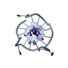
- PDB-6pnk: Crystal structure of the G-quadruplex formed by (GGGTT)3GGG in co... -
+ Open data

- Basic information
| Entry | Database: PDB / ID: 6pnk | ||||||||||||||||||||||||||||
|---|---|---|---|---|---|---|---|---|---|---|---|---|---|---|---|---|---|---|---|---|---|---|---|---|---|---|---|---|---|
| Title | Crystal structure of the G-quadruplex formed by (GGGTT)3GGG in complex with N-methylmesoporphryin IX | ||||||||||||||||||||||||||||
 Components Components | DNA (5'-D(P* Keywords KeywordsDNA / parallel G-quadruplex / end-stacking / TT propeller loops | Function / homology | FORMIC ACID / : / N-METHYLMESOPORPHYRIN / DNA / DNA (> 10) |  Function and homology information Function and homology informationBiological species |   Tetrahymena thermophila (eukaryote) Tetrahymena thermophila (eukaryote)Method |  X-RAY DIFFRACTION / SYNCHROTRON / MOLECULAR REPLACEMENT / Resolution: 2.39 Å X-RAY DIFFRACTION / SYNCHROTRON / MOLECULAR REPLACEMENT / Resolution: 2.39 Å  Authors AuthorsYatsunyk, L.A. / Lin, L.Y. | Funding support | |  United States, 1items United States, 1items
 CitationJournal: Plos One / Year: 2020 CitationJournal: Plos One / Year: 2020Title: Biophysical and X-ray structural studies of the (GGGTT)3GGG G-quadruplex in complex with N-methyl mesoporphyrin IX. Authors: Lin, L.Y. / McCarthy, S. / Powell, B.M. / Manurung, Y. / Xiang, I.M. / Dean, W.L. / Chaires, B. / Yatsunyk, L.A. History |
|
- Structure visualization
| Structure viewer | Molecule: MolmilJmol/JSmol |
|---|
- Downloads & links
-Download
| PDBx/mmCIF format | 6pnk.cif.gz | 66.8 KB | Display | PDBx/mmCIF format |
|---|---|---|---|---|
| PDB format | pdb6pnk.ent.gz | 42.8 KB | Display | PDB format |
| PDBx/mmJSON format | 6pnk.json.gz | Tree view | PDBx/mmJSON format | |
| Others |  Other downloads Other downloads |
-Validation report
| Arichive directory | https://data.pdbj.org/pub/pdb/validation_reports/pn/6pnkftp://data.pdbj.org/pub/pdb/validation_reports/pn/6pnk | HTTPS FTP |
|---|
-Related structure data
| Related structure data | 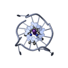 6p45SC S: Starting model for refinement C: citing same article ( |
|---|---|
| Similar structure data |


-Links
 PDBj
PDBj


- Assembly
| Deposited unit | 
| ||||||||||||
|---|---|---|---|---|---|---|---|---|---|---|---|---|---|
| 1 | 
| ||||||||||||
| 2 | 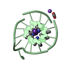
| ||||||||||||
| Unit cell |
| ||||||||||||
| Components on special symmetry positions |
|
-Components
| #1: DNA chain | Mass: 5730.669 Da / Num. of mol.: 2 / Source method: obtained synthetically / Source: (synth.) Tetrahymena thermophila (eukaryote)#2: Chemical | ChemComp-K /   Mass: 39.098 Da / Num. of mol.: 6 / Source method: obtained synthetically / Formula: K Mass: 39.098 Da / Num. of mol.: 6 / Source method: obtained synthetically / Formula: K#3: Chemical | 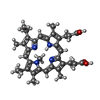  Mass: 580.716 Da / Num. of mol.: 2 / Source method: obtained synthetically / Formula: C35H40N4O4 / Feature type: SUBJECT OF INVESTIGATION Mass: 580.716 Da / Num. of mol.: 2 / Source method: obtained synthetically / Formula: C35H40N4O4 / Feature type: SUBJECT OF INVESTIGATION#4: Chemical | ChemComp-FMT / |   Mass: 46.025 Da / Num. of mol.: 1 / Source method: obtained synthetically / Formula: CH2O2 Mass: 46.025 Da / Num. of mol.: 1 / Source method: obtained synthetically / Formula: CH2O2#5: Chemical | ChemComp-NA / |   Mass: 22.990 Da / Num. of mol.: 1 / Source method: obtained synthetically / Formula: Na Mass: 22.990 Da / Num. of mol.: 1 / Source method: obtained synthetically / Formula: NaHas ligand of interest | Y | |
|---|
-Experimental details
-Experiment
| Experiment | Method: X-RAY DIFFRACTION / Number of used crystals: 1 |
|---|
- Sample preparation
| Crystal | Density Matthews: 3.03 Å3/Da / Density % sol: 59.39 % / Description: Hexagonal |
|---|---|
| Crystal grow | Temperature: 293 K / Method: vapor diffusion, hanging drop / pH: 7 Details: 0.85 M sodium formate, 17.5% PEG 20000, 0.05 M Bis-Tris pH 7.0 |
-Data collection
| Diffraction | Mean temperature: 196 K / Serial crystal experiment: N |
|---|---|
| Diffraction source | Source: SYNCHROTRON / Site: APS / Beamline: 24-ID-C / Wavelength: 0.9791 Å |
| Detector | Type: DECTRIS PILATUS 6M-F / Detector: PIXEL / Date: Mar 10, 2018 |
| Radiation | Monochromator: Cryo-Cooled double crystal / Protocol: SINGLE WAVELENGTH / Monochromatic (M) / Laue (L): M / Scattering type: x-ray |
| Radiation wavelength | Wavelength: 0.9791 Å / Relative weight: 1 |
| Reflection | Resolution: 2.388→64.78 Å / Num. obs: 5748 / % possible obs: 97.5 % / Redundancy: 19 % / Biso Wilson estimate: 90.97 Å2 / CC1/2: 0.369 / Rmerge(I) obs: 0.067 / Net I/σ(I): 21.4 |
| Reflection shell | Resolution: 2.388→2.473 Å / Redundancy: 19 % / Mean I/σ(I) obs: 0.6 / Num. unique obs: 492 / CC1/2: 0.369 / % possible all: 86.9 |
- Processing
| Software |
| ||||||||||||||||||||||||||||||||||||||||
|---|---|---|---|---|---|---|---|---|---|---|---|---|---|---|---|---|---|---|---|---|---|---|---|---|---|---|---|---|---|---|---|---|---|---|---|---|---|---|---|---|---|
| Refinement | Method to determine structure: MOLECULAR REPLACEMENT Starting model: 6P45 Resolution: 2.39→64.78 Å / SU ML: 0.2982 / Cross valid method: FREE R-VALUE / σ(F): 1.35 / Phase error: 37.4204
| ||||||||||||||||||||||||||||||||||||||||
| Solvent computation | Shrinkage radii: 0.9 Å / VDW probe radii: 1.11 Å | ||||||||||||||||||||||||||||||||||||||||
| Displacement parameters | Biso mean: 115.44 Å2 | ||||||||||||||||||||||||||||||||||||||||
| Refinement step | Cycle: LAST / Resolution: 2.39→64.78 Å
| ||||||||||||||||||||||||||||||||||||||||
| Refine LS restraints |
| ||||||||||||||||||||||||||||||||||||||||
| LS refinement shell |
| ||||||||||||||||||||||||||||||||||||||||
| Refinement TLS params. | Method: refined / Origin x: -15.9855156741 Å / Origin y: 8.42790186784 Å / Origin z: -48.7943706657 Å
| ||||||||||||||||||||||||||||||||||||||||
| Refinement TLS group | Selection details: all |
