Movie
Movie Controller


+ Open data

- Basic information
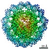
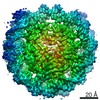
| Entry | Database: EMDB / ID: EMD-4768 | ||||||||||||||||||||||||
|---|---|---|---|---|---|---|---|---|---|---|---|---|---|---|---|---|---|---|---|---|---|---|---|---|---|
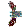
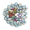
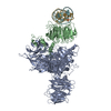
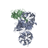
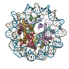
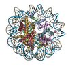
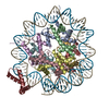
| Title | Cryo-EM structure of NCP_THF2(-3) | ||||||||||||||||||||||||
 Map data Map data | |||||||||||||||||||||||||
 Sample Sample |
| ||||||||||||||||||||||||
 Keywords Keywords | DNA damage / Nucleosome / 6-4 photoproduct / DNA BINDING PROTEIN | ||||||||||||||||||||||||
| Function / homology |  Function and homology information Function and homology informationnegative regulation of tumor necrosis factor-mediated signaling pathway / negative regulation of megakaryocyte differentiation / protein localization to CENP-A containing chromatin / Chromatin modifying enzymes / Replacement of protamines by nucleosomes in the male pronucleus / CENP-A containing nucleosome / Packaging Of Telomere Ends / Recognition and association of DNA glycosylase with site containing an affected purine / Cleavage of the damaged purine / Recognition and association of DNA glycosylase with site containing an affected pyrimidine ...negative regulation of tumor necrosis factor-mediated signaling pathway / negative regulation of megakaryocyte differentiation / protein localization to CENP-A containing chromatin / Chromatin modifying enzymes / Replacement of protamines by nucleosomes in the male pronucleus / CENP-A containing nucleosome / Packaging Of Telomere Ends / Recognition and association of DNA glycosylase with site containing an affected purine / Cleavage of the damaged purine / Recognition and association of DNA glycosylase with site containing an affected pyrimidine / Cleavage of the damaged pyrimidine / Deposition of new CENPA-containing nucleosomes at the centromere / telomere organization / Interleukin-7 signaling / Inhibition of DNA recombination at telomere / Meiotic synapsis / RNA Polymerase I Promoter Opening / Assembly of the ORC complex at the origin of replication / innate immune response in mucosa / Regulation of endogenous retroelements by the Human Silencing Hub (HUSH) complex / SUMOylation of chromatin organization proteins / DNA methylation / Condensation of Prophase Chromosomes / Chromatin modifications during the maternal to zygotic transition (MZT) / HCMV Late Events / SIRT1 negatively regulates rRNA expression / epigenetic regulation of gene expression / ERCC6 (CSB) and EHMT2 (G9a) positively regulate rRNA expression / PRC2 methylates histones and DNA / Regulation of endogenous retroelements by KRAB-ZFP proteins / Defective pyroptosis / Regulation of endogenous retroelements by Piwi-interacting RNAs (piRNAs) / HDACs deacetylate histones / Nonhomologous End-Joining (NHEJ) / RNA Polymerase I Promoter Escape / lipopolysaccharide binding / Transcriptional regulation by small RNAs / Formation of the beta-catenin:TCF transactivating complex / RUNX1 regulates genes involved in megakaryocyte differentiation and platelet function / Activated PKN1 stimulates transcription of AR (androgen receptor) regulated genes KLK2 and KLK3 / HDMs demethylate histones / G2/M DNA damage checkpoint / NoRC negatively regulates rRNA expression / DNA Damage/Telomere Stress Induced Senescence / B-WICH complex positively regulates rRNA expression / PKMTs methylate histone lysines / Meiotic recombination / Pre-NOTCH Transcription and Translation / Metalloprotease DUBs / RMTs methylate histone arginines / Activation of anterior HOX genes in hindbrain development during early embryogenesis / Transcriptional regulation of granulopoiesis / antimicrobial humoral immune response mediated by antimicrobial peptide / HCMV Early Events / structural constituent of chromatin / antibacterial humoral response / UCH proteinases / nucleosome / heterochromatin formation / E3 ubiquitin ligases ubiquitinate target proteins / nucleosome assembly / Recruitment and ATM-mediated phosphorylation of repair and signaling proteins at DNA double strand breaks / HATs acetylate histones / RUNX1 regulates transcription of genes involved in differentiation of HSCs / chromatin organization / Factors involved in megakaryocyte development and platelet production / MLL4 and MLL3 complexes regulate expression of PPARG target genes in adipogenesis and hepatic steatosis / Processing of DNA double-strand break ends / Senescence-Associated Secretory Phenotype (SASP) / defense response to Gram-negative bacterium / Oxidative Stress Induced Senescence / killing of cells of another organism / gene expression / Estrogen-dependent gene expression / chromosome, telomeric region / defense response to Gram-positive bacterium / Ub-specific processing proteases / cadherin binding / Amyloid fiber formation / protein heterodimerization activity / negative regulation of cell population proliferation / protein-containing complex / DNA binding / extracellular space / RNA binding / extracellular exosome / extracellular region / nucleoplasm / nucleus / membrane / cytosol Similarity search - Function | ||||||||||||||||||||||||
| Biological species |  Homo sapiens (human) Homo sapiens (human) | ||||||||||||||||||||||||
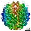
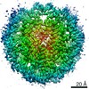
| Method | single particle reconstruction / cryo EM / Resolution: 3.5 Å | ||||||||||||||||||||||||
 Authors Authors | Matsumoto S / Cavadini S | ||||||||||||||||||||||||
| Funding support |  Switzerland, Switzerland,  Japan, 7 items Japan, 7 items
| ||||||||||||||||||||||||
 Citation Citation | Journal: Nature / Year: 2019 Title: DNA damage detection in nucleosomes involves DNA register shifting. Authors: Syota Matsumoto / Simone Cavadini / Richard D Bunker / Ralph S Grand / Alessandro Potenza / Julius Rabl / Junpei Yamamoto / Andreas D Schenk / Dirk Schübeler / Shigenori Iwai / Kaoru ...Authors: Syota Matsumoto / Simone Cavadini / Richard D Bunker / Ralph S Grand / Alessandro Potenza / Julius Rabl / Junpei Yamamoto / Andreas D Schenk / Dirk Schübeler / Shigenori Iwai / Kaoru Sugasawa / Hitoshi Kurumizaka / Nicolas H Thomä / Abstract: Access to DNA packaged in nucleosomes is critical for gene regulation, DNA replication and DNA repair. In humans, the UV-damaged DNA-binding protein (UV-DDB) complex detects UV-light-induced ...Access to DNA packaged in nucleosomes is critical for gene regulation, DNA replication and DNA repair. In humans, the UV-damaged DNA-binding protein (UV-DDB) complex detects UV-light-induced pyrimidine dimers throughout the genome; however, it remains unknown how these lesions are recognized in chromatin, in which nucleosomes restrict access to DNA. Here we report cryo-electron microscopy structures of UV-DDB bound to nucleosomes bearing a 6-4 pyrimidine-pyrimidone dimer or a DNA-damage mimic in various positions. We find that UV-DDB binds UV-damaged nucleosomes at lesions located in the solvent-facing minor groove without affecting the overall nucleosome architecture. In the case of buried lesions that face the histone core, UV-DDB changes the predominant translational register of the nucleosome and selectively binds the lesion in an accessible, exposed position. Our findings explain how UV-DDB detects occluded lesions in strongly positioned nucleosomes, and identify slide-assisted site exposure as a mechanism by which high-affinity DNA-binding proteins can access otherwise occluded sites in nucleosomal DNA. | ||||||||||||||||||||||||
| History |
|
- Structure visualization
| Movie |
Movie viewer |
|---|---|
| Structure viewer | EM map: SurfViewMolmilJmol/JSmol |
| Supplemental images |

- Downloads & links
-EMDB archive
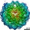
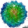
| Map data | emd_4768.map.gz | 19.7 MB | EMDB map data format | |
|---|---|---|---|---|
| Header (meta data) | emd-4768-v30.xmlemd-4768.xml | 25.3 KB 25.3 KB | Display Display | EMDB header |
| Images | 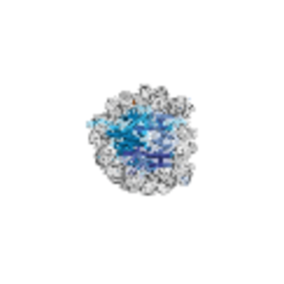 emd_4768.png emd_4768.png | 125.5 KB | ||
| Filedesc metadata | emd-4768.cif.gz | 7.1 KB | ||
| Archive directory |  http://ftp.pdbj.org/pub/emdb/structures/EMD-4768ftp://ftp.pdbj.org/pub/emdb/structures/EMD-4768 http://ftp.pdbj.org/pub/emdb/structures/EMD-4768ftp://ftp.pdbj.org/pub/emdb/structures/EMD-4768 | HTTPS FTP |
-Validation report
| Summary document | emd_4768_validation.pdf.gz | 385 KB | Display | EMDB validaton report |
|---|---|---|---|---|
| Full document | emd_4768_full_validation.pdf.gz | 384.6 KB | Display | |
| Data in XML | emd_4768_validation.xml.gz | 6.3 KB | Display | |
| Data in CIF | emd_4768_validation.cif.gz | 7.2 KB | Display | |
| Arichive directory | https://ftp.pdbj.org/pub/emdb/validation_reports/EMD-4768ftp://ftp.pdbj.org/pub/emdb/validation_reports/EMD-4768 | HTTPS FTP |
-Related structure data
| Related structure data | 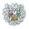 6r94MC 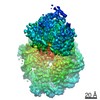 4762C 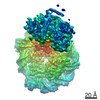 4763C 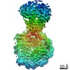 4764C 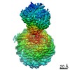 4765C 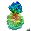 4766C 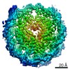 4767C 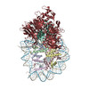 6r8yC  6r8zC 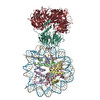 6r90C 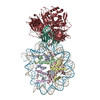 6r91C 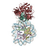 6r92C 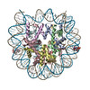 6r93C M: atomic model generated by this map C: citing same article ( |
|---|---|
| Similar structure data |

-Links
| EMDB pages | EMDB (EBI/PDBe) / EMDataResource |
|---|---|
| Related items in Molecule of the Month |


-Map
| File | Download / File: emd_4768.map.gz / Format: CCP4 / Size: 103 MB / Type: IMAGE STORED AS FLOATING POINT NUMBER (4 BYTES) | ||||||||||||||||||||||||||||||||||||||||||||||||||||||||||||||||||||
|---|---|---|---|---|---|---|---|---|---|---|---|---|---|---|---|---|---|---|---|---|---|---|---|---|---|---|---|---|---|---|---|---|---|---|---|---|---|---|---|---|---|---|---|---|---|---|---|---|---|---|---|---|---|---|---|---|---|---|---|---|---|---|---|---|---|---|---|---|---|
| Projections & slices | Image control
Images are generated by Spider. | ||||||||||||||||||||||||||||||||||||||||||||||||||||||||||||||||||||
| Voxel size | X=Y=Z: 0.86 Å | ||||||||||||||||||||||||||||||||||||||||||||||||||||||||||||||||||||
| Density |
| ||||||||||||||||||||||||||||||||||||||||||||||||||||||||||||||||||||
| Symmetry | Space group: 1 | ||||||||||||||||||||||||||||||||||||||||||||||||||||||||||||||||||||
| Details | EMDB XML:
CCP4 map header:
| ||||||||||||||||||||||||||||||||||||||||||||||||||||||||||||||||||||
 Z (Sec.)
 Y (Row.)
 X (Col.)


-Supplemental data
- Sample components
+Entire : THF2 (-3) containing nucleosome
+Supramolecule #1: THF2 (-3) containing nucleosome
+Supramolecule #2: Human DNA
+Supramolecule #3: Histone H4
+Supramolecule #4: Histone H3.1 / 2A / 2B
+Macromolecule #1: Human alpha-satellite DNA (145-MER)
+Macromolecule #2: Human alpha-satellite DNA (145-MER) with abasic sites at position...
+Macromolecule #3: Histone H3.1

+Macromolecule #4: Histone H4
+Macromolecule #5: Histone H2A type 1-B/E
+Macromolecule #6: Histone H2B type 1-J
-Experimental details
-Structure determination
| Method | cryo EM |
|---|---|
 Processing Processing | single particle reconstruction |
| Aggregation state | particle |
-Sample preparation
| Concentration | 2 mg/mL | ||||||||||||
|---|---|---|---|---|---|---|---|---|---|---|---|---|---|
| Buffer | pH: 7.4 Component:
| ||||||||||||
| Vitrification | Cryogen name: ETHANE / Chamber humidity: 85 % / Chamber temperature: 277 K / Instrument: LEICA EM GP |
- Electron microscopy
| Microscope | FEI TITAN KRIOS |
|---|---|
| Specialist optics | Energy filter - Name: GIF Quantum LS / Energy filter - Slit width: 20 eV |
| Image recording | Film or detector model: GATAN K2 SUMMIT (4k x 4k) / Detector mode: COUNTING / Number grids imaged: 1 / Average exposure time: 6.0 sec. / Average electron dose: 40.0 e/Å2 |
| Electron beam | Acceleration voltage: 300 kV / Electron source:  FIELD EMISSION GUN FIELD EMISSION GUN |
| Electron optics | C2 aperture diameter: 50.0 µm / Illumination mode: SPOT SCAN / Imaging mode: BRIGHT FIELD / Cs: 0.0 mm / Nominal magnification: 130000 |
| Sample stage | Specimen holder model: FEI TITAN KRIOS AUTOGRID HOLDER / Cooling holder cryogen: NITROGEN |
| Experimental equipment |  Model: Titan Krios / Image courtesy: FEI Company |
-Image processing
| Startup model | Type of model: OTHER |
|---|---|
| Final reconstruction | Resolution.type: BY AUTHOR / Resolution: 3.5 Å / Resolution method: FSC 0.143 CUT-OFF / Software - Name: RELION (ver. 2) / Number images used: 78672 |
| Initial angle assignment | Type: OTHER / Software - Name: RELION |
| Final angle assignment | Type: OTHER / Software - Name: RELION |
-Atomic model buiding 1
| Initial model |
| ||||||||||||||||||||||||||
|---|---|---|---|---|---|---|---|---|---|---|---|---|---|---|---|---|---|---|---|---|---|---|---|---|---|---|---|
| Refinement | Space: REAL / Protocol: FLEXIBLE FIT / Target criteria: Cross-correlation coefficient | ||||||||||||||||||||||||||
| Output model | PDB-6r94: |