 Movie
Movie Controller

[English] 日本語
 Yorodumi
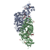
- PDB-4e54: Damaged DNA induced UV-damaged DNA-binding protein (UV-DDB) dimer... -
+ Open data

- Basic information
| Entry | Database: PDB / ID: 4.0E+54 | ||||||
|---|---|---|---|---|---|---|---|
| Title | Damaged DNA induced UV-damaged DNA-binding protein (UV-DDB) dimerization and its roles in chromatinized DNA repair | ||||||
 Components Components |
| ||||||
 Keywords Keywords | DNA BINDING PROTEIN/DNA / BETA BARREL / DOUBLE HELIX / DDB1:WD40 beta-barrel fold / DNA damage / DNA repair / Host-virus interactions / Protein ubiquitination / Proteosomal degradation / DNA BINDING PROTEIN-DNA complex | ||||||
| Function / homology |  Function and homology information Function and homology informationpositive regulation by virus of viral protein levels in host cell / spindle assembly involved in female meiosis / epigenetic programming in the zygotic pronuclei / UV-damage excision repair / biological process involved in interaction with symbiont / regulation of mitotic cell cycle phase transition / WD40-repeat domain binding / Cul4A-RING E3 ubiquitin ligase complex / Cul4-RING E3 ubiquitin ligase complex / Cul4B-RING E3 ubiquitin ligase complex ...positive regulation by virus of viral protein levels in host cell / spindle assembly involved in female meiosis / epigenetic programming in the zygotic pronuclei / UV-damage excision repair / biological process involved in interaction with symbiont / regulation of mitotic cell cycle phase transition / WD40-repeat domain binding / Cul4A-RING E3 ubiquitin ligase complex / Cul4-RING E3 ubiquitin ligase complex / Cul4B-RING E3 ubiquitin ligase complex / ubiquitin ligase complex scaffold activity / negative regulation of reproductive process / negative regulation of developmental process / pyrimidine dimer repair / viral release from host cell / cullin family protein binding / ectopic germ cell programmed cell death / response to UV / positive regulation of viral genome replication / protein autoubiquitination / site of DNA damage / proteasomal protein catabolic process / positive regulation of gluconeogenesis / TP53 Regulates Transcription of DNA Repair Genes / nucleotide-excision repair / sperm end piece / regulation of circadian rhythm / Recognition of DNA damage by PCNA-containing replication complex / DNA Damage Recognition in GG-NER / Dual Incision in GG-NER / Wnt signaling pathway / Transcription-Coupled Nucleotide Excision Repair (TC-NER) / Formation of TC-NER Pre-Incision Complex / Formation of Incision Complex in GG-NER / protein polyubiquitination / Dual incision in TC-NER / positive regulation of protein catabolic process / Gap-filling DNA repair synthesis and ligation in TC-NER / cellular response to UV / cell junction / rhythmic process / site of double-strand break / sperm principal piece / Neddylation / sperm midpiece / ubiquitin-dependent protein catabolic process / damaged DNA binding / proteasome-mediated ubiquitin-dependent protein catabolic process / protein-macromolecule adaptor activity / chromosome, telomeric region / Ub-specific processing proteases / protein ubiquitination / DNA repair / apoptotic process / DNA damage response / negative regulation of apoptotic process / chromatin / protein-containing complex binding / nucleolus / protein-containing complex / : / DNA binding / extracellular exosome / nucleoplasm / nucleus / cytoplasm Similarity search - Function | ||||||
| Biological species |  Homo sapiens (human) Homo sapiens (human) | ||||||
| Method |  X-RAY DIFFRACTION / SYNCHROTRON / MOLECULAR REPLACEMENT / Resolution: 2.85 Å X-RAY DIFFRACTION / SYNCHROTRON / MOLECULAR REPLACEMENT / Resolution: 2.85 Å | ||||||
 Authors Authors | Yeh, J.I. / Du, S. | ||||||
 Citation Citation | Journal: Proc.Natl.Acad.Sci.USA / Year: 2012 Title: Damaged DNA induced UV-damaged DNA-binding protein (UV-DDB) dimerization and its roles in chromatinized DNA repair. Authors: Yeh, J.I. / Levine, A.S. / Du, S. / Chinte, U. / Ghodke, H. / Wang, H. / Shi, H. / Hsieh, C.L. / Conway, J.F. / Van Houten, B. / Rapic-Otrin, V. | ||||||
| History |
|
- Structure visualization
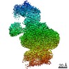
| Structure viewer | Molecule: MolmilJmol/JSmol |
|---|
- Downloads & links
-Download
| PDBx/mmCIF format | 4e54.cif.gz | 626.4 KB | Display | PDBx/mmCIF format |
|---|---|---|---|---|
| PDB format | pdb4e54.ent.gz | 495.9 KB | Display | PDB format |
| PDBx/mmJSON format | 4e54.json.gz | Tree view | PDBx/mmJSON format | |
| Others |  Other downloads Other downloads |
-Validation report
| Arichive directory | https://data.pdbj.org/pub/pdb/validation_reports/e5/4e54ftp://data.pdbj.org/pub/pdb/validation_reports/e5/4e54 | HTTPS FTP |
|---|
-Related structure data
| Related structure data | 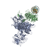 4e5zC 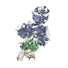 3ei2S S: Starting model for refinement C: citing same article ( |
|---|---|
| Similar structure data |


-Links
 PDBj
PDBj


- Assembly
| Deposited unit | 
| ||||||||
|---|---|---|---|---|---|---|---|---|---|
| 1 |
| ||||||||
| Unit cell |
|
-Components
| #1: Protein | Mass: 128478.914 Da / Num. of mol.: 1 / Fragment: DNA DAMAGE-BINDING PROTEIN 1 (DDB1; p127) / Mutation: NT-His10-DDB1 Source method: isolated from a genetically manipulated source Source: (gene. exp.) Homo sapiens (human) / Gene: DDB1, DDB1_HUMAN, Q16531, XAP1 / Plasmid: pBlueBac4.5/V5-His NT-His10-DDB1 / Cell line (production host): Sf9 / Production host:   Spodoptera frugiperda (fall armyworm) / References: UniProt: Q16531 Spodoptera frugiperda (fall armyworm) / References: UniProt: Q16531 |
|---|---|
| #2: Protein | Mass: 48928.906 Da / Num. of mol.: 1 / Fragment: DNA DAMAGE-BINDING PROTEIN 2 (DDB2; p48) / Mutation: N-FLAG-DDB2 Source method: isolated from a genetically manipulated source Source: (gene. exp.) Homo sapiens (human) / Gene: DDB2, DDB2_HUMAN, Q92466 / Plasmid: pBlueBac4.5/V5-His NT-FLAG-DDB2 / Cell line (production host): Sf9 / Production host: Spodoptera frugiperda (fall armyworm) / References: UniProt: Q92466 |
| #3: DNA chain | Mass: 7424.801 Da / Num. of mol.: 1 / Fragment: AP24 DNA strand / Source method: obtained synthetically Details: Synthetic single stranded 24-oligodeoxynucleotides with complementary strand sequence: 5-TGACTGTATGATGACGATGCTGAC-3 |
| #4: DNA chain | Mass: 7189.646 Da / Num. of mol.: 1 / Fragment: AP24 DNA complementary strand / Source method: obtained synthetically Details: Synthetic single stranded oligodeoxynucleotides with a central tetrahydrofuran abasic site mimic (3DR) on coding strand with sequence: 5-GTCAGCATCG(3DR)CATCATACAGTCA-3 |
| Has protein modification | Y |
-Experimental details
-Experiment
| Experiment | Method: X-RAY DIFFRACTION / Number of used crystals: 1 |
|---|
- Sample preparation
| Crystal | Density Matthews: 2.67 Å3/Da / Density % sol: 53.95 % |
|---|---|
| Crystal grow | Temperature: 277 K / Method: vapor diffusion / pH: 7.5 Details: 20mM Tris pH 7.5, 2mM MgCl2, 1mM EDTA, 2mM TECP, 5% Glycerol, 0.02% azide. UV-DDB-AP24 complex (molar ratio of 1:3 UV-DDB:DNA) at 2.5 mg/mL. 'AP24' refers to synthetic DNA substrate of 24- ...Details: 20mM Tris pH 7.5, 2mM MgCl2, 1mM EDTA, 2mM TECP, 5% Glycerol, 0.02% azide. UV-DDB-AP24 complex (molar ratio of 1:3 UV-DDB:DNA) at 2.5 mg/mL. 'AP24' refers to synthetic DNA substrate of 24-bpr with a central abasic site mimic., VAPOR DIFFUSION, temperature 277K |
-Data collection
| Diffraction | Mean temperature: 100 K |
|---|---|
| Diffraction source | Source: SYNCHROTRON / Site: APS  / Beamline: 22-ID / Wavelength: 1 Å / Beamline: 22-ID / Wavelength: 1 Å |
| Detector | Type: MARMOSAIC 300 mm CCD / Detector: CCD / Date: Jul 8, 2009 / Details: monochromators |
| Radiation | Monochromator: SAGITALLY FOCUSED Si(111) / Protocol: SINGLE WAVELENGTH / Monochromatic (M) / Laue (L): M / Scattering type: x-ray |
| Radiation wavelength | Wavelength: 1 Å / Relative weight: 1 |
| Reflection | Resolution: 2.85→50 Å / Num. obs: 47108 / % possible obs: 89.7 % / Observed criterion σ(F): 2 / Observed criterion σ(I): 2 / Redundancy: 6.5 % / Biso Wilson estimate: 25.22 Å2 / Rmerge(I) obs: 0.12 / Rsym value: 0.1 / Net I/σ(I): 12.28 |
| Reflection shell | Resolution: 2.85→2.96 Å / Redundancy: 3.5 % / Rmerge(I) obs: 0.12 / Mean I/σ(I) obs: 7.7 / Num. unique all: 3545 / Rsym value: 0.321 / % possible all: 67.6 |
- Processing
| Software |
| ||||||||||||||||||||||||||||||||||||||||||||||||||||||||||||||||||||||||||||||||||||||||||||||||||||||||||||||||||||||||||||||||||||||||||||||||||||||
|---|---|---|---|---|---|---|---|---|---|---|---|---|---|---|---|---|---|---|---|---|---|---|---|---|---|---|---|---|---|---|---|---|---|---|---|---|---|---|---|---|---|---|---|---|---|---|---|---|---|---|---|---|---|---|---|---|---|---|---|---|---|---|---|---|---|---|---|---|---|---|---|---|---|---|---|---|---|---|---|---|---|---|---|---|---|---|---|---|---|---|---|---|---|---|---|---|---|---|---|---|---|---|---|---|---|---|---|---|---|---|---|---|---|---|---|---|---|---|---|---|---|---|---|---|---|---|---|---|---|---|---|---|---|---|---|---|---|---|---|---|---|---|---|---|---|---|---|---|---|---|---|
| Refinement | Method to determine structure: MOLECULAR REPLACEMENT Starting model: PDB Entry 3EI2 Resolution: 2.85→33.315 Å / SU ML: 0.79 / Isotropic thermal model: Isotropic / Cross valid method: THROUGHOUT / σ(F): 1.34 / σ(I): 2.7 / Phase error: 24.09 / Stereochemistry target values: MLHL
| ||||||||||||||||||||||||||||||||||||||||||||||||||||||||||||||||||||||||||||||||||||||||||||||||||||||||||||||||||||||||||||||||||||||||||||||||||||||
| Solvent computation | Shrinkage radii: 0.77 Å / VDW probe radii: 0.9 Å / Solvent model: FLAT BULK SOLVENT MODEL / Bsol: 299.989 Å2 / ksol: 0.265 e/Å3 | ||||||||||||||||||||||||||||||||||||||||||||||||||||||||||||||||||||||||||||||||||||||||||||||||||||||||||||||||||||||||||||||||||||||||||||||||||||||
| Displacement parameters |
| ||||||||||||||||||||||||||||||||||||||||||||||||||||||||||||||||||||||||||||||||||||||||||||||||||||||||||||||||||||||||||||||||||||||||||||||||||||||
| Refine analyze | Luzzati sigma a free: 0.367 Å | ||||||||||||||||||||||||||||||||||||||||||||||||||||||||||||||||||||||||||||||||||||||||||||||||||||||||||||||||||||||||||||||||||||||||||||||||||||||
| Refinement step | Cycle: LAST / Resolution: 2.85→33.315 Å
| ||||||||||||||||||||||||||||||||||||||||||||||||||||||||||||||||||||||||||||||||||||||||||||||||||||||||||||||||||||||||||||||||||||||||||||||||||||||
| Refine LS restraints |
| ||||||||||||||||||||||||||||||||||||||||||||||||||||||||||||||||||||||||||||||||||||||||||||||||||||||||||||||||||||||||||||||||||||||||||||||||||||||
| LS refinement shell |
| ||||||||||||||||||||||||||||||||||||||||||||||||||||||||||||||||||||||||||||||||||||||||||||||||||||||||||||||||||||||||||||||||||||||||||||||||||||||
| Refinement TLS params. | Method: refined / Refine-ID: X-RAY DIFFRACTION
| ||||||||||||||||||||||||||||||||||||||||||||||||||||||||||||||||||||||||||||||||||||||||||||||||||||||||||||||||||||||||||||||||||||||||||||||||||||||
| Refinement TLS group |
|
