 Movie
Movie Controller

[English] 日本語
 Yorodumi
- PDB-4ci3: Structure of the DDB1-CRBN E3 ubiquitin ligase bound to Pomalidomide -
+ Open data

- Basic information
| Entry | Database: PDB / ID: 4ci3 | ||||||
|---|---|---|---|---|---|---|---|
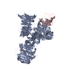
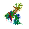
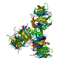
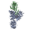
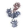
| Title | Structure of the DDB1-CRBN E3 ubiquitin ligase bound to Pomalidomide | ||||||
 Components Components |
| ||||||
 Keywords Keywords | DNA BINDING PROTEIN / DDB1 / CRBN / CULLIN / E3 LIGASE / UBIQUITIN / THALIDOMIDE / CONTERGAN | ||||||
| Function / homology |  Function and homology information Function and homology informationpositive regulation by virus of viral protein levels in host cell / spindle assembly involved in female meiosis / epigenetic programming in the zygotic pronuclei / UV-damage excision repair / biological process involved in interaction with symbiont / limb development / regulation of mitotic cell cycle phase transition / WD40-repeat domain binding / Cul4A-RING E3 ubiquitin ligase complex / Cul4-RING E3 ubiquitin ligase complex ...positive regulation by virus of viral protein levels in host cell / spindle assembly involved in female meiosis / epigenetic programming in the zygotic pronuclei / UV-damage excision repair / biological process involved in interaction with symbiont / limb development / regulation of mitotic cell cycle phase transition / WD40-repeat domain binding / Cul4A-RING E3 ubiquitin ligase complex / Cul4-RING E3 ubiquitin ligase complex / Cul4B-RING E3 ubiquitin ligase complex / ubiquitin ligase complex scaffold activity / negative regulation of reproductive process / negative regulation of developmental process / viral release from host cell / cullin family protein binding / ectopic germ cell programmed cell death / positive regulation of viral genome replication / positive regulation of Wnt signaling pathway / proteasomal protein catabolic process / positive regulation of gluconeogenesis / nucleotide-excision repair / sperm end piece / regulation of circadian rhythm / Recognition of DNA damage by PCNA-containing replication complex / DNA Damage Recognition in GG-NER / Dual Incision in GG-NER / Wnt signaling pathway / Transcription-Coupled Nucleotide Excision Repair (TC-NER) / Formation of TC-NER Pre-Incision Complex / Formation of Incision Complex in GG-NER / Dual incision in TC-NER / positive regulation of protein catabolic process / Gap-filling DNA repair synthesis and ligation in TC-NER / cellular response to UV / rhythmic process / site of double-strand break / sperm principal piece / Neddylation / sperm midpiece / ubiquitin-dependent protein catabolic process / damaged DNA binding / proteasome-mediated ubiquitin-dependent protein catabolic process / protein-macromolecule adaptor activity / chromosome, telomeric region / protein ubiquitination / DNA repair / apoptotic process / DNA damage response / negative regulation of apoptotic process / protein-containing complex binding / nucleolus / protein-containing complex / : / DNA binding / extracellular exosome / nucleoplasm / metal ion binding / nucleus / cytoplasm Similarity search - Function | ||||||
| Biological species |  HOMO SAPIENS (human) HOMO SAPIENS (human) | ||||||
| Method |  X-RAY DIFFRACTION / SYNCHROTRON / MOLECULAR REPLACEMENT / Resolution: 3.5 Å X-RAY DIFFRACTION / SYNCHROTRON / MOLECULAR REPLACEMENT / Resolution: 3.5 Å | ||||||
 Authors Authors | Fischer, E.S. / Boehm, K. / Thoma, N.H. | ||||||
 Citation Citation | Journal: Nature / Year: 2014 Title: Structure of the Ddb1-Crbn E3 Ubiquitin Ligase in Complex with Thalidomide. Authors: Fischer, E.S. / Bohm, K. / Lydeard, J.R. / Yang, H. / Stadler, M.B. / Cavadini, S. / Nagel, J. / Serluca, F. / Acker, V. / Lingaraju, G.M. / Tichkule, R.B. / Schebesta, M. / Forrester, W.C. ...Authors: Fischer, E.S. / Bohm, K. / Lydeard, J.R. / Yang, H. / Stadler, M.B. / Cavadini, S. / Nagel, J. / Serluca, F. / Acker, V. / Lingaraju, G.M. / Tichkule, R.B. / Schebesta, M. / Forrester, W.C. / Schirle, M. / Hassiepen, U. / Ottl, J. / Hild, M. / Beckwith, R.E.J. / Harper, J.W. / Jenkins, J.L. / Thoma, N.H. | ||||||
| History |
|
- Structure visualization
| Structure viewer | Molecule: MolmilJmol/JSmol |
|---|
- Downloads & links
-Download
| PDBx/mmCIF format | 4ci3.cif.gz | 596.3 KB | Display | PDBx/mmCIF format |
|---|---|---|---|---|
| PDB format | pdb4ci3.ent.gz | 489.8 KB | Display | PDB format |
| PDBx/mmJSON format | 4ci3.json.gz | Tree view | PDBx/mmJSON format | |
| Others |  Other downloads Other downloads |
-Validation report
| Arichive directory | https://data.pdbj.org/pub/pdb/validation_reports/ci/4ci3ftp://data.pdbj.org/pub/pdb/validation_reports/ci/4ci3 | HTTPS FTP |
|---|
-Related structure data
| Related structure data |  4ci1C  4ci2C 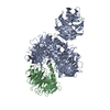 3ei3S C: citing same article ( S: Starting model for refinement |
|---|---|
| Similar structure data |


-Links
 PDBj
PDBj


- Assembly
| Deposited unit | 
| ||||||||
|---|---|---|---|---|---|---|---|---|---|
| 1 |
| ||||||||
| Unit cell |
|
-Components
| #1: Protein | Mass: 129253.898 Da / Num. of mol.: 1 Source method: isolated from a genetically manipulated source Source: (gene. exp.) HOMO SAPIENS (human) / Plasmid: PFASTBAC DUAL / Cell line (production host): High Five / Production host:  TRICHOPLUSIA NI (cabbage looper) / References: UniProt: Q16531 TRICHOPLUSIA NI (cabbage looper) / References: UniProt: Q16531 |
|---|---|
| #2: Protein | Mass: 53967.180 Da / Num. of mol.: 1 Source method: isolated from a genetically manipulated source Source: (gene. exp.) TRICHOPLUSIA NI (cabbage looper) / References: UniProt: P0CF65 |
| #3: Chemical | ChemComp-ZN /   Mass: 65.409 Da / Num. of mol.: 1 / Source method: obtained synthetically / Formula: Zn Mass: 65.409 Da / Num. of mol.: 1 / Source method: obtained synthetically / Formula: Zn |
| #4: Chemical | ChemComp-Y70 /   Mass: 273.244 Da / Num. of mol.: 1 / Source method: obtained synthetically / Formula: C13H11N3O4 Mass: 273.244 Da / Num. of mol.: 1 / Source method: obtained synthetically / Formula: C13H11N3O4 |
-Experimental details
-Experiment
| Experiment | Method: X-RAY DIFFRACTION / Number of used crystals: 1 |
|---|
- Sample preparation
| Crystal | Density Matthews: 3.51 Å3/Da / Density % sol: 65 % / Description: NONE |
|---|---|
| Crystal grow | pH: 6.5 Details: PROTEIN WAS CRYSTALLIZED FROM 100 MM NA-CACOCYLATE, 80 MM NAH2PO4, 120 MM K2HPO4, 700 MM TRI-NA CITRATE., pH 6.5 |
-Data collection
| Diffraction | Mean temperature: 100 K |
|---|---|
| Diffraction source | Source: SYNCHROTRON / Site: SLS  / Beamline: X10SA / Wavelength: 0.99997 / Beamline: X10SA / Wavelength: 0.99997 |
| Detector | Type: DECTRIS PILATUS 6M / Detector: PIXEL / Date: Nov 14, 2012 / Details: MIRRORS |
| Radiation | Monochromator: SI(111) MONOCHROMATOR / Protocol: SINGLE WAVELENGTH / Monochromatic (M) / Laue (L): M / Scattering type: x-ray |
| Radiation wavelength | Wavelength: 0.99997 Å / Relative weight: 1 |
| Reflection | Resolution: 3.5→30 Å / Num. obs: 25109 / % possible obs: 84.4 % / Observed criterion σ(I): -3 / Redundancy: 2.6 % / Biso Wilson estimate: 88.2 Å2 / Rmerge(I) obs: 0.14 / Net I/σ(I): 7.05 |
| Reflection shell | Resolution: 3.5→3.59 Å / Redundancy: 2.5 % / Rmerge(I) obs: 1.31 / Mean I/σ(I) obs: 0.91 / % possible all: 83.4 |
- Processing
| Software |
| ||||||||||||||||||||||||||||||||||||||||||||||||||||||||||||||||||||||||||||||||||||||||||||||||||||||||||||||||||
|---|---|---|---|---|---|---|---|---|---|---|---|---|---|---|---|---|---|---|---|---|---|---|---|---|---|---|---|---|---|---|---|---|---|---|---|---|---|---|---|---|---|---|---|---|---|---|---|---|---|---|---|---|---|---|---|---|---|---|---|---|---|---|---|---|---|---|---|---|---|---|---|---|---|---|---|---|---|---|---|---|---|---|---|---|---|---|---|---|---|---|---|---|---|---|---|---|---|---|---|---|---|---|---|---|---|---|---|---|---|---|---|---|---|---|---|
| Refinement | Method to determine structure: MOLECULAR REPLACEMENT Starting model: PDB ENTRY 3EI3, CHAIN A Resolution: 3.5→29.63 Å / Cor.coef. Fo:Fc: 0.9254 / Cor.coef. Fo:Fc free: 0.9159 / Cross valid method: THROUGHOUT / σ(F): 0 / SU Rfree Blow DPI: 0.567
| ||||||||||||||||||||||||||||||||||||||||||||||||||||||||||||||||||||||||||||||||||||||||||||||||||||||||||||||||||
| Displacement parameters | Biso mean: 139.22 Å2
| ||||||||||||||||||||||||||||||||||||||||||||||||||||||||||||||||||||||||||||||||||||||||||||||||||||||||||||||||||
| Refine analyze | Luzzati coordinate error obs: 0.921 Å | ||||||||||||||||||||||||||||||||||||||||||||||||||||||||||||||||||||||||||||||||||||||||||||||||||||||||||||||||||
| Refinement step | Cycle: LAST / Resolution: 3.5→29.63 Å
| ||||||||||||||||||||||||||||||||||||||||||||||||||||||||||||||||||||||||||||||||||||||||||||||||||||||||||||||||||
| Refine LS restraints |
| ||||||||||||||||||||||||||||||||||||||||||||||||||||||||||||||||||||||||||||||||||||||||||||||||||||||||||||||||||
| LS refinement shell | Resolution: 3.5→3.64 Å / Total num. of bins used: 13
| ||||||||||||||||||||||||||||||||||||||||||||||||||||||||||||||||||||||||||||||||||||||||||||||||||||||||||||||||||
| Refinement TLS params. | Method: refined / Refine-ID: X-RAY DIFFRACTION
|
