[English] 日本語
Yorodumi
- PDB-4tz4: Crystal Structure of Human Cereblon in Complex with DDB1 and Lena... -

+
Open data


ID or keywords:

Loading...

-
Basic information

Entry
Database: PDB / ID: 4tz4
Title
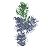
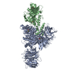
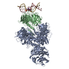
Crystal Structure of Human Cereblon in Complex with DDB1 and Lenalidomide
Components
  • DNA damage-binding protein 1
  • Protein cereblon
KeywordsDNA Binding Protein/Ligase / DCAF / DNA Binding Protein-Ligase complex
Function / homology
Function and homology information


negative regulation of monoatomic ion transmembrane transport / positive regulation by virus of viral protein levels in host cell / epigenetic programming in the zygotic pronuclei / spindle assembly involved in female meiosis / Cul4-RING E3 ubiquitin ligase complex / UV-damage excision repair / biological process involved in interaction with symbiont / regulation of mitotic cell cycle phase transition / WD40-repeat domain binding / Cul4A-RING E3 ubiquitin ligase complex ...negative regulation of monoatomic ion transmembrane transport / positive regulation by virus of viral protein levels in host cell / epigenetic programming in the zygotic pronuclei / spindle assembly involved in female meiosis / Cul4-RING E3 ubiquitin ligase complex / UV-damage excision repair / biological process involved in interaction with symbiont / regulation of mitotic cell cycle phase transition / WD40-repeat domain binding / Cul4A-RING E3 ubiquitin ligase complex / Cul4B-RING E3 ubiquitin ligase complex / ubiquitin ligase complex scaffold activity / locomotory exploration behavior / negative regulation of reproductive process / negative regulation of developmental process / cullin family protein binding / positive regulation of Wnt signaling pathway / viral release from host cell / ectopic germ cell programmed cell death / positive regulation of viral genome replication / negative regulation of protein-containing complex assembly / positive regulation of gluconeogenesis / proteasomal protein catabolic process / Recognition of DNA damage by PCNA-containing replication complex / nucleotide-excision repair / positive regulation of protein-containing complex assembly / DNA Damage Recognition in GG-NER / Dual Incision in GG-NER / regulation of circadian rhythm / Transcription-Coupled Nucleotide Excision Repair (TC-NER) / Formation of TC-NER Pre-Incision Complex / Wnt signaling pathway / Formation of Incision Complex in GG-NER / Dual incision in TC-NER / Gap-filling DNA repair synthesis and ligation in TC-NER / positive regulation of protein catabolic process / cellular response to UV / rhythmic process / protein-macromolecule adaptor activity / site of double-strand break / Neddylation / ubiquitin-dependent protein catabolic process / proteasome-mediated ubiquitin-dependent protein catabolic process / Potential therapeutics for SARS / transmembrane transporter binding / damaged DNA binding / chromosome, telomeric region / protein ubiquitination / DNA repair / apoptotic process / DNA damage response / protein-containing complex binding / nucleolus / negative regulation of apoptotic process / perinuclear region of cytoplasm / protein-containing complex / DNA binding / extracellular space / extracellular exosome / nucleoplasm / membrane / metal ion binding / nucleus / cytosol / cytoplasm
Similarity search - Function
Methane Monooxygenase Hydroxylase; Chain G, domain 1 - #1480 / LON domain-like / Peptide methionine sulfoxide reductase. / DNA polymerase; domain 1 - #910 / Yippee/Mis18/Cereblon / Yippee zinc-binding/DNA-binding /Mis18, centromere assembly / Metal Binding Protein, Guanine Nucleotide Exchange Factor; Chain A / Archaeosine Trna-guanine Transglycosylase; Chain: A, domain 4 / CULT domain / CULT domain profile. ...Methane Monooxygenase Hydroxylase; Chain G, domain 1 - #1480 / LON domain-like / Peptide methionine sulfoxide reductase. / DNA polymerase; domain 1 - #910 / Yippee/Mis18/Cereblon / Yippee zinc-binding/DNA-binding /Mis18, centromere assembly / Metal Binding Protein, Guanine Nucleotide Exchange Factor; Chain A / Archaeosine Trna-guanine Transglycosylase; Chain: A, domain 4 / CULT domain / CULT domain profile. / Lon protease, N-terminal domain superfamily / Lon N-terminal domain profile. / Lon protease, N-terminal domain / ATP-dependent protease La (LON) substrate-binding domain / Found in ATP-dependent protease La (LON) / Cleavage/polyadenylation specificity factor, A subunit, N-terminal / Mono-functional DNA-alkylating methyl methanesulfonate N-term / Cleavage/polyadenylation specificity factor, A subunit, C-terminal / CPSF A subunit region / PUA-like superfamily / YVTN repeat-like/Quinoprotein amine dehydrogenase / 7 Propeller / Methylamine Dehydrogenase; Chain H / Methane Monooxygenase Hydroxylase; Chain G, domain 1 / Beta Complex / DNA polymerase; domain 1 / Roll / WD40-repeat-containing domain superfamily / WD40/YVTN repeat-like-containing domain superfamily / Up-down Bundle / Orthogonal Bundle / Mainly Beta / Mainly Alpha
Similarity search - Domain/homology
S-Lenalidomide / DNA damage-binding protein 1 / Protein cereblon
Similarity search - Component
Biological speciesHomo sapiens (human)
MethodX-RAY DIFFRACTION / SYNCHROTRON / MOLECULAR REPLACEMENT / Resolution: 3.01 Å
AuthorsChamberlain, P.P. / Pagarigan, B. / Delker, S. / Leon, B. / Riley, M.
CitationJournal: To Be Published
Title: Structural Basis for Responsiveness to Thalidomide-Analog Drugs Defined by the Crystal Structure of the Human Cereblon:DDB1:Lenalidomide Complex
Authors: Chamberlain, P.P. / Wang, M. / Riley, M. / Delker, S. / Carmel, G. / Miller, K. / Lopez-Girona, A. / Pagarigan, B. / Leon, B. / Rychak, E. / Corral, L. / Lopez-Girona, A. / Ren, Y. / Wang, M. ...Authors: Chamberlain, P.P. / Wang, M. / Riley, M. / Delker, S. / Carmel, G. / Miller, K. / Lopez-Girona, A. / Pagarigan, B. / Leon, B. / Rychak, E. / Corral, L. / Lopez-Girona, A. / Ren, Y. / Wang, M. / Riley, M. / Delker, S. / Ito, T. / Hideki, A. / Mori, T. / Handa, H. / Hakoshima, T. / Daniel, T.O. / Miller, K. / Cathers, B.E. / Carmel, G. / Pagarigan, B. / Leon, B. / Rychak, E. / Corral, L. / Ren, Y.
History
DepositionJul 9, 2014Deposition site: RCSB / Processing site: RCSB
Revision 1.0Aug 6, 2014Provider: repository / Type: Initial release
Revision 2.0Dec 27, 2023Group: Atomic model / Data collection ...Atomic model / Data collection / Database references / Derived calculations / Other / Refinement description / Source and taxonomy
Category: atom_site_anisotrop / chem_comp_atom ...atom_site_anisotrop / chem_comp_atom / chem_comp_bond / citation / database_2 / entity_src_gen / pdbx_database_status / pdbx_struct_oper_list / refine_hist
Item: _atom_site_anisotrop.pdbx_PDB_model_num / _atom_site_anisotrop.pdbx_label_asym_id ..._atom_site_anisotrop.pdbx_PDB_model_num / _atom_site_anisotrop.pdbx_label_asym_id / _atom_site_anisotrop.pdbx_label_atom_id / _atom_site_anisotrop.pdbx_label_comp_id / _atom_site_anisotrop.pdbx_label_seq_id / _citation.journal_id_CSD / _database_2.pdbx_DOI / _database_2.pdbx_database_accession / _entity_src_gen.pdbx_alt_source_flag / _pdbx_database_status.pdb_format_compatible / _pdbx_struct_oper_list.symmetry_operation / _refine_hist.pdbx_number_atoms_nucleic_acid / _refine_hist.pdbx_number_atoms_protein
Revision 2.1Apr 3, 2024Group: Refinement description / Category: pdbx_initial_refinement_model

-
Structure visualization

Structure viewerMolecule:
MolmilJmol/JSmol

Downloads & links

-
Assembly

Deposited unit
A: DNA damage-binding protein 1
C: Protein cereblon
hetero molecules


Theoretical massNumber of molelcules
Total (without water)172,0714
Polymers171,7472
Non-polymers3252
Water32418
1


  • Idetical with deposited unit
  • defined by author&software
TypeNameSymmetry operationNumber
identity operation1_555x,y,z1
Buried area4320 Å2
ΔGint-25 kcal/mol
Surface area59790 Å2
MethodPISA
Unit cell
Length a, b, c (Å)71.862, 129.117, 198.677
Angle α, β, γ (deg.)90.000, 90.000, 90.000
Int Tables number19
Space group name H-MP212121
Detailsbiological unit is the same as asym.

-
Components

#1: Protein DNA damage-binding protein 1 / DDB p127 subunit / DNA damage-binding protein a / DDBa / Damage-specific DNA-binding protein 1 / ...DDB p127 subunit / DNA damage-binding protein a / DDBa / Damage-specific DNA-binding protein 1 / HBV X-associated protein 1 / XAP-1 / UV-damaged DNA-binding factor / UV-damaged DNA-binding protein 1 / UV-DDB 1 / XPE-binding factor / XPE-BF / Xeroderma pigmentosum group E-complementing protein / XPCe


Mass: 127879.219 Da / Num. of mol.: 1
Source method: isolated from a genetically manipulated source
Source: (gene. exp.) Homo sapiens (human) / Gene: DDB1, XAP1 / Production host: 
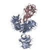
Spodoptera frugiperda (fall armyworm) / References: UniProt: Q16531
#2: Protein Protein cereblon


Mass: 43867.391 Da / Num. of mol.: 1
Source method: isolated from a genetically manipulated source
Source: (gene. exp.) Homo sapiens (human) / Gene: CRBN, AD-006 / Production host: Spodoptera frugiperda (fall armyworm) / References: UniProt: Q96SW2
#3: Chemical ChemComp-ZN / ZINC ION


Mass: 65.409 Da / Num. of mol.: 1 / Source method: obtained synthetically / Formula: Zn
#4: Chemical ChemComp-LVY / S-Lenalidomide


Mass: 259.261 Da / Num. of mol.: 1 / Source method: obtained synthetically / Formula: C13H13N3O3
#5: Water ChemComp-HOH / water / Water


Mass: 18.015 Da / Num. of mol.: 18 / Source method: isolated from a natural source / Formula: H2O

-
Experimental details

-
Experiment

ExperimentMethod: X-RAY DIFFRACTION / Number of used crystals: 1

-
Sample preparation

CrystalDensity Matthews: 2.68 Å3/Da / Density % sol: 54.16 %
Crystal growTemperature: 293 K / Method: vapor diffusion, sitting drop / pH: 7.5 / Details: 100mM HEPES pH 7.5, 18% PEG 10K

-
Data collection

DiffractionMean temperature: 100 K
Diffraction sourceSource: SYNCHROTRON / Site: CLSI / Beamline: 08ID-1 / Wavelength: 1 Å
DetectorType: MARMOSAIC 300 mm CCD / Detector: CCD / Date: Jan 15, 2014
RadiationProtocol: SINGLE WAVELENGTH / Monochromatic (M) / Laue (L): M / Scattering type: x-ray
Radiation wavelengthWavelength: 1 Å / Relative weight: 1
ReflectionResolution: 3.01→50 Å / Num. obs: 37313 / % possible obs: 98.5 % / Redundancy: 6.1 % / Rmerge(I) obs: 0.181 / Χ2: 1.058 / Net I/av σ(I): 10.061 / Net I/σ(I): 5.9 / Num. measured all: 228575
Reflection shell

Diffraction-ID: 1 / Rejects: 0

Resolution (Å)Redundancy (%)Rmerge(I) obsNum. unique allΧ2% possible all
3.01-3.125.80.72331841.0285.8
3.12-3.246.30.58437031.08299.9
3.24-3.396.30.39237311.08100
3.39-3.576.30.29437391.054100
3.57-3.796.30.2337681.045100
3.79-4.086.20.18737451.04599.9
4.08-4.56.20.14737951.034100
4.5-5.156.10.12137961.08899.9
5.15-6.486.10.13638351.08799.9
6.48-505.60.08240171.03699.5

-
Processing

Software
NameVersionClassification
REFMAC5.6.0117refinement
HKL-3000data reduction
PDB_EXTRACT3.14data extraction
Cootmodel building
RefinementMethod to determine structure: MOLECULAR REPLACEMENT
Starting model: DDB1

Resolution: 3.01→50 Å / Cor.coef. Fo:Fc: 0.918 / Cor.coef. Fo:Fc free: 0.873 / SU B: 38.571 / SU ML: 0.328 / Cross valid method: THROUGHOUT / σ(F): 0 / ESU R Free: 0.465 / Stereochemistry target values: MAXIMUM LIKELIHOOD
Details: HYDROGENS HAVE BEEN USED IF PRESENT IN THE INPUT U VALUES : RESIDUAL ONLY
RfactorNum. reflection% reflectionSelection details
Rfree0.2706 1857 5 %RANDOM
Rwork0.2015 35396 --
obs0.2049 37253 99.4 %-
Solvent computationIon probe radii: 0.8 Å / Shrinkage radii: 0.8 Å / VDW probe radii: 1.2 Å / Solvent model: MASK
Displacement parametersBiso max: 79.43 Å2 / Biso mean: 30.294 Å2 / Biso min: 11.62 Å2
Baniso -1Baniso -2Baniso -3
1--1.52 Å20 Å20 Å2
2--1.67 Å20 Å2
3----0.16 Å2
Refinement stepCycle: final / Resolution: 3.01→50 Å
ProteinNucleic acidLigandSolventTotal
Num. atoms11043 0 20 18 11081
Biso mean--38.05 25.48 -
Num. residues----1431
Refine LS restraints
Refine-IDTypeDev idealDev ideal targetNumber
X-RAY DIFFRACTIONr_bond_refined_d0.0080.0211307
X-RAY DIFFRACTIONr_angle_refined_deg1.2111.9615349
X-RAY DIFFRACTIONr_dihedral_angle_1_deg7.38251410
X-RAY DIFFRACTIONr_dihedral_angle_2_deg39.81224.444477
X-RAY DIFFRACTIONr_dihedral_angle_3_deg19.871151876
X-RAY DIFFRACTIONr_dihedral_angle_4_deg21.1841553
X-RAY DIFFRACTIONr_chiral_restr0.0820.21786
X-RAY DIFFRACTIONr_gen_planes_refined0.0080.0218412
LS refinement shellResolution: 3.01→3.088 Å / Total num. of bins used: 20
RfactorNum. reflection% reflection
Rfree0.342 111 -
Rwork0.27 2223 -
all-2334 -
obs--92.95 %
Refinement TLS params.

Method: refined / Refine-ID: X-RAY DIFFRACTION

IDL112)L122)L132)L222)L232)L332)S11 (Å °)S12 (Å °)S13 (Å °)S21 (Å °)S22 (Å °)S23 (Å °)S31 (Å °)S32 (Å °)S33 (Å °)T112)T122)T132)T222)T232)T332)Origin x (Å)Origin y (Å)Origin z (Å)
13.58141.04380.20933.2779-0.40013.21250.1427-0.36940.35310.414-0.1791-0.0495-0.480.52610.03630.1819-0.1725-0.02230.34010.00730.0512-0.295515.1173-7.6299
20.6744-0.086-0.12851.59280.40010.94170.01390.0822-0.0329-0.217-0.014-0.08570.06280.01730.00010.1202-0.01180.00690.11550.0450.0815-13.09043.8364-65.2819
31.1981-0.3355-0.16310.69110.00810.6488-0.026-0.02450.1280.06160.03970.0221-0.0573-0.0277-0.01370.1081-0.01710.03110.06760.01680.1493-21.449729.8526-47.2447
40.2447-0.85380.27454.0544-0.75721.5630.1740.0776-0.0597-0.1843-0.01330.20170.4191-0.0262-0.16070.31120.0353-0.06290.21680.01880.1573-31.494919.5893-66.7327
51.245-0.55520.87091.2852-0.87881.94470.0184-0.08270.04080.0163-0.1457-0.0825-0.063-0.00840.12730.21440.0164-0.01120.11390.0240.1062-36.929247.0136-87.0997
Refinement TLS group
IDRefine-IDRefine TLS-IDAuth asym-IDAuth seq-ID
1X-RAY DIFFRACTION1A393 - 708
2X-RAY DIFFRACTION2A2 - 392
3X-RAY DIFFRACTION3A709 - 1147
4X-RAY DIFFRACTION4C187 - 256
5X-RAY DIFFRACTION5C48 - 186
6X-RAY DIFFRACTION5C257 - 428

+
About Yorodumi

-
News

-
Feb 9, 2022. New format data for meta-information of EMDB entries

New format data for meta-information of EMDB entries

  • Version 3 of the EMDB header file is now the official format.
  • The previous official version 1.9 will be removed from the archive.

Related info.:EMDB header

External links:wwPDB to switch to version 3 of the EMDB data model

-
Aug 12, 2020. Covid-19 info

Covid-19 info

URL: https://pdbj.org/emnavi/covid19.php

New page: Covid-19 featured information page in EM Navigator.

Related info.:Covid-19 info / Mar 5, 2020. Novel coronavirus structure data

+
Mar 5, 2020. Novel coronavirus structure data

Novel coronavirus structure data

Related info.:Yorodumi Speices / Aug 12, 2020. Covid-19 info

External links:COVID-19 featured content - PDBj / Molecule of the Month (242):Coronavirus Proteases

+
Jan 31, 2019. EMDB accession codes are about to change! (news from PDBe EMDB page)

EMDB accession codes are about to change! (news from PDBe EMDB page)

  • The allocation of 4 digits for EMDB accession codes will soon come to an end. Whilst these codes will remain in use, new EMDB accession codes will include an additional digit and will expand incrementally as the available range of codes is exhausted. The current 4-digit format prefixed with “EMD-” (i.e. EMD-XXXX) will advance to a 5-digit format (i.e. EMD-XXXXX), and so on. It is currently estimated that the 4-digit codes will be depleted around Spring 2019, at which point the 5-digit format will come into force.
  • The EM Navigator/Yorodumi systems omit the EMD- prefix.

Related info.:Q: What is EMD? / ID/Accession-code notation in Yorodumi/EM Navigator

External links:EMDB Accession Codes are Changing Soon! / Contact to PDBj

+
Jul 12, 2017. Major update of PDB

Major update of PDB

  • wwPDB released updated PDB data conforming to the new PDBx/mmCIF dictionary.
  • This is a major update changing the version number from 4 to 5, and with Remediation, in which all the entries are updated.
  • In this update, many items about electron microscopy experimental information are reorganized (e.g. em_software).
  • Now, EM Navigator and Yorodumi are based on the updated data.

External links:wwPDB Remediation / Enriched Model Files Conforming to OneDep Data Standards Now Available in the PDB FTP Archive

-
Yorodumi

Thousand views of thousand structures

  • Yorodumi is a browser for structure data from EMDB, PDB, SASBDB, etc.
  • This page is also the successor to EM Navigator detail page, and also detail information page/front-end page for Omokage search.
  • The word "yorodu" (or yorozu) is an old Japanese word meaning "ten thousand". "mi" (miru) is to see.

Related info.:EMDB / PDB / SASBDB / Comparison of 3 databanks / Yorodumi Search / Aug 31, 2016. New EM Navigator & Yorodumi / Yorodumi Papers / Jmol/JSmol / Function and homology information / Changes in new EM Navigator and Yorodumi

Read more