DNA BINDING PROTEIN/DNA / UV-damage / DDB / nucleotide excision repair / xeroderma pigmentosum / Alternative splicing / Disease mutation / DNA damage / DNA repair / DNA-binding / Nucleus / Phosphoprotein / Polymorphism / WD repeat / Ubl conjugation pathway / DNA BINDING PROTEIN-DNA COMPLEX
Function / homology
Function and homology information
Dual Incision in GG-NER / DNA Damage Recognition in GG-NER / Formation of Incision Complex in GG-NER / Neddylation / positive regulation by virus of viral protein levels in host cell / spindle assembly involved in female meiosis / epigenetic programming in the zygotic pronuclei / UV-damage excision repair / biological process involved in interaction with symbiont / regulation of mitotic cell cycle phase transition ...Dual Incision in GG-NER / DNA Damage Recognition in GG-NER / Formation of Incision Complex in GG-NER / Neddylation / positive regulation by virus of viral protein levels in host cell / spindle assembly involved in female meiosis / epigenetic programming in the zygotic pronuclei / UV-damage excision repair / biological process involved in interaction with symbiont / regulation of mitotic cell cycle phase transition / WD40-repeat domain binding / Cul4A-RING E3 ubiquitin ligase complex / Cul4-RING E3 ubiquitin ligase complex / Cul4B-RING E3 ubiquitin ligase complex / ubiquitin ligase complex scaffold activity / negative regulation of reproductive process / negative regulation of developmental process / viral release from host cell / cullin family protein binding / ectopic germ cell programmed cell death / response to UV / positive regulation of viral genome replication / site of DNA damage / proteasomal protein catabolic process / positive regulation of gluconeogenesis / nucleotide-excision repair / sperm end piece / regulation of circadian rhythm / Recognition of DNA damage by PCNA-containing replication complex / DNA Damage Recognition in GG-NER / Dual Incision in GG-NER / Wnt signaling pathway / Transcription-Coupled Nucleotide Excision Repair (TC-NER) / Formation of TC-NER Pre-Incision Complex / Formation of Incision Complex in GG-NER / Dual incision in TC-NER / positive regulation of protein catabolic process / Gap-filling DNA repair synthesis and ligation in TC-NER / cellular response to UV / rhythmic process / site of double-strand break / sperm principal piece / Neddylation / sperm midpiece / ubiquitin-dependent protein catabolic process / damaged DNA binding / proteasome-mediated ubiquitin-dependent protein catabolic process / protein-macromolecule adaptor activity / chromosome, telomeric region / protein ubiquitination / DNA repair / apoptotic process / DNA damage response / negative regulation of apoptotic process / protein-containing complex binding / nucleolus / protein-containing complex / : / DNA binding / extracellular exosome / nucleoplasm / nucleus / cytoplasm Similarity search - Function
Helix Hairpins - #3280 / DNA damage-binding protein 2 / DNA polymerase; domain 1 - #910 / : / RSE1/DDB1/CPSF1 second beta-propeller / Cleavage/polyadenylation specificity factor, A subunit, C-terminal / Cleavage/polyadenylation specificity factor, A subunit, N-terminal / : / CPSF A subunit region / RSE1/DDB1/CPSF1 first beta-propeller ...Helix Hairpins - #3280 / DNA damage-binding protein 2 / DNA polymerase; domain 1 - #910 / : / RSE1/DDB1/CPSF1 second beta-propeller / Cleavage/polyadenylation specificity factor, A subunit, C-terminal / Cleavage/polyadenylation specificity factor, A subunit, N-terminal / : / CPSF A subunit region / RSE1/DDB1/CPSF1 first beta-propeller / YVTN repeat-like/Quinoprotein amine dehydrogenase / 7 Propeller / Methylamine Dehydrogenase; Chain H / DNA polymerase; domain 1 / Helix Hairpins / WD domain, G-beta repeat / WD40 repeat, conserved site / Trp-Asp (WD) repeats signature. / Trp-Asp (WD) repeats profile. / Trp-Asp (WD) repeats circular profile. / WD40 repeats / WD40 repeat / WD40-repeat-containing domain superfamily / WD40/YVTN repeat-like-containing domain superfamily / Orthogonal Bundle / Mainly Beta / Mainly Alpha Similarity search - Domain/homology
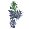
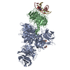
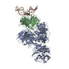
A: DNA damage-binding protein 1 B: DNA damage-binding protein 2 G: 5'-D(*DAP*DCP*DGP*DCP*DGP*DAP*(64T)P*(5PY)P*DGP*DCP*DGP*DCP*DCP*DC)-3' H: 5'-D(*DTP*DGP*DGP*DGP*DCP*DGP*DCP*DAP*DAP*DTP*DCP*DGP*DCP*DG)-3' hetero molecules
DNA damage-binding protein ... , 2 types, 2 molecules AB
#1: Protein
DNAdamage-bindingprotein1 / hsDDB1 / Damage-specific DNA-binding protein 1 / UV-damaged DNA-binding factor / DDB p127 subunit / ...hsDDB1 / Damage-specific DNA-binding protein 1 / UV-damaged DNA-binding factor / DDB p127 subunit / DNA damage-binding protein a / DDBa / UV-damaged DNA-binding protein 1 / UV-DDB 1 / Xeroderma pigmentosum group E-complementing protein / XPCe / XPE-binding factor / XPE-BF / HBV X-associated protein 1 / XAP-1
Mass: 129253.898 Da / Num. of mol.: 1 Source method: isolated from a genetically manipulated source Source: (gene. exp.) Homo sapiens (human) / Gene: DDB1, XAP1 / Cell (production host): high five cells / Cell line (production host): BTI-TN-5B1-4 / Production host: Trichoplusia ni (cabbage looper) / References: UniProt: Q16531
#2: Protein
DNAdamage-bindingprotein2 / Damage-specific DNA-binding protein 2
Mass: 43559.113 Da / Num. of mol.: 1 Source method: isolated from a genetically manipulated source Source: (gene. exp.) Danio rerio (zebrafish) / Gene: ddb2, si:dkey-45f10.3 / Cell (production host): high five cells / Cell line (production host): BTI-TN-5B1-4 / Production host: Trichoplusia ni (cabbage looper) / References: UniProt: Q2YDS1
In the structure databanks used in Yorodumi, some data are registered as the other names, "COVID-19 virus" and "2019-nCoV". Here are the details of the virus and the list of structure data.
Jan 31, 2019. EMDB accession codes are about to change! (news from PDBe EMDB page)
EMDB accession codes are about to change! (news from PDBe EMDB page)
The allocation of 4 digits for EMDB accession codes will soon come to an end. Whilst these codes will remain in use, new EMDB accession codes will include an additional digit and will expand incrementally as the available range of codes is exhausted. The current 4-digit format prefixed with “EMD-” (i.e. EMD-XXXX) will advance to a 5-digit format (i.e. EMD-XXXXX), and so on. It is currently estimated that the 4-digit codes will be depleted around Spring 2019, at which point the 5-digit format will come into force.
The EM Navigator/Yorodumi systems omit the EMD- prefix.
Related info.:Q: What is EMD? / ID/Accession-code notation in Yorodumi/EM Navigator
Yorodumi is a browser for structure data from EMDB, PDB, SASBDB, etc.
This page is also the successor to EM Navigator detail page, and also detail information page/front-end page for Omokage search.
The word "yorodu" (or yorozu) is an old Japanese word meaning "ten thousand". "mi" (miru) is to see.
Related info.:EMDB / PDB / SASBDB / Comparison of 3 databanks / Yorodumi Search / Aug 31, 2016. New EM Navigator & Yorodumi / Yorodumi Papers / Jmol/JSmol / Function and homology information / Changes in new EM Navigator and Yorodumi
 Movie
Movie Controller

 Yorodumi
 Open data
 Basic information
 Components
 Keywords
 Function and homology information
 Homo sapiens (human)
 X-RAY DIFFRACTION /
  Authors
 Citation
 Structure visualization
 Downloads & links
 Other downloads


 PDBj
PDBj


 Assembly
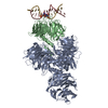
 Trichoplusia ni (cabbage looper) / References: UniProt: Q16531

 Mass: 194.226 Da / Num. of mol.: 1 / Source method: obtained synthetically / Formula: C8H18O5 / Comment: precipitant*YM
 Sample preparation
 / Beamline: X10SA / Wavelength: 0.9918 Å
 Processing