Protocol: SINGLE WAVELENGTH / Monochromatic (M) / Laue (L): M / Scattering type: x-ray
Radiation wavelength
Wavelength: 1 Å / Relative weight: 1
Reflection
Resolution: 1.88→50 Å / Num. obs: 76864 / % possible obs: 99.9 % / Redundancy: 22.8 % / Rmerge(I) obs: 0.089 / Χ2: 1.019 / Net I/av σ(I): 38.415 / Net I/σ(I): 9.7 / Num. measured all: 1750335
Reflection shell
Diffraction-ID: 1 / Rejects: _
Resolution (Å)
Redundancy (%)
Rmerge(I) obs
Num. unique all
Χ2
% possible all
1.88-1.95
23
0.599
7652
1.049
100
1.95-2.03
23.1
0.367
7739
1.012
100
2.03-2.12
23.2
0.278
7642
1.002
100
2.12-2.23
22.8
0.213
7671
1.042
100
2.23-2.37
20.5
0.174
7747
1.006
100
2.37-2.55
23.4
0.147
7687
1.008
100
2.55-2.81
23.5
0.105
7678
1.009
100
2.81-3.21
23.5
0.077
7684
1.025
100
3.21-4.05
21.5
0.067
7661
1.018
99.5
4.05-50
23.3
0.049
7703
1.018
100
-
Processing
Software
Name
Version
Classification
REFMAC
5.6.0117
refinement
HKL-2000
datareduction
PDB_EXTRACT
3.14
dataextraction
DENZO
datareduction
SCALEPACK
datascaling
Refinement
Resolution: 1.88→50 Å / Cor.coef. Fo:Fc: 0.965 / Cor.coef. Fo:Fc free: 0.943 / SU B: 5.102 / SU ML: 0.085 / Cross valid method: THROUGHOUT / ESU R: 0.13 / ESU R Free: 0.13 / Stereochemistry target values: MAXIMUM LIKELIHOOD / Details: HYDROGENS HAVE BEEN USED IF PRESENT IN THE INPUT
Rfactor
Num. reflection
% reflection
Selection details
Rfree
0.21973
1983
5 %
RANDOM
Rwork
0.17289
-
-
-
obs
0.1752
37584
99.94 %
-
Solvent computation
Ion probe radii: 0.8 Å / Shrinkage radii: 0.8 Å / VDW probe radii: 1.2 Å / Solvent model: MASK
 Movie
Movie Controller


 Open data
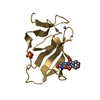
 Basic information
 Components
 Keywords
 Function and homology information
 X-RAY DIFFRACTION /
  Authors
 Citation
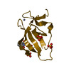
 Structure visualization
 Downloads & links
 Other downloads
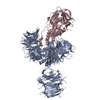


 PDBj
PDBj
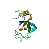
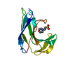
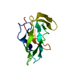
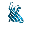
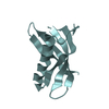
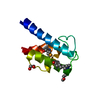
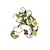
 Assembly
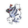


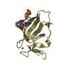
 Mass: 65.409 Da / Num. of mol.: 4 / Source method: obtained synthetically / Formula: Zn
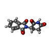
 Mass: 258.229 Da / Num. of mol.: 4 / Source method: obtained synthetically / Formula: C13H10N2O4 / Comment: medication*YM
 Mass: 96.063 Da / Num. of mol.: 8 / Source method: obtained synthetically / Formula: SO4
 Mass: 18.015 Da / Num. of mol.: 365 / Source method: isolated from a natural source / Formula: H2O
 Sample preparation
 / Beamline: 22-ID / Wavelength: 1 Å
 Processing