D res high: 2.1 Å / D res low: 50 Å / FOM : 0.273 / FOM acentric: 0.31 / FOM centric: 0 / Reflection: 8041 / Reflection acentric: 7089 / Reflection centric: 952
Phasing MAD set
R cullis acentric: 1.54 / R cullis centric: 1 / Highest resolution: 2.1 Å / Lowest resolution: 50 Å / Loc acentric: 0.1 / Loc centric: 0.1 / Power acentric: 0 / Power centric: 0 / Reflection acentric: 7089 / Reflection centric: 952
Phasing MAD set shell
ID: 1 / R cullis centric: 1 / Power acentric: 0 / Power centric: 0
Resolution (Å)
R cullis acentric
Loc acentric
Loc centric
Reflection acentric
Reflection centric
12.98-50
1.56
0.4
0.2
22
15
7.46-12.98
1.05
0.3
0.2
107
49
5.23-7.46
1.26
0.2
0.1
287
79
4.03-5.23
1.16
0.2
0.1
530
101
3.28-4.03
1.14
0.1
0.1
858
137
2.76-3.28
1.94
0
0
1270
157
2.39-2.76
5.08
0
0
1756
200
2.1-2.39
7.77
0
0
2259
214
Phasing MAD set site
Atom type symbol: Se / B iso: 63.7551 / Fract x: -0.34 / Fract y: -0.506 / Fract z: -0.007 / Occupancy: 5.185 / Occupancy iso: 0
Phasing MAD shell
Resolution (Å)
FOM
FOM acentric
FOM centric
Reflection
Reflection acentric
Reflection centric
12.98-50
0.226
0.381
0
37
22
15
7.46-12.98
0.313
0.457
0
156
107
49
5.23-7.46
0.408
0.52
0
366
287
79
4.03-5.23
0.403
0.479
0
631
530
101
3.28-4.03
0.412
0.478
0
995
858
137
2.76-3.28
0.398
0.447
0
1427
1270
157
2.39-2.76
0.255
0.284
0
1956
1756
200
2.1-2.39
0.106
0.116
0
2473
2259
214
Phasing dm
Method: Solvent flattening and Histogram matching / Reflection: 8041
Phasing dm shell
Resolution (Å)
Delta phi final
FOM
Reflection
5.42-100
64.4
0.826
501
4.27-5.42
63.4
0.914
504
3.72-4.27
59.3
0.925
505
3.37-3.72
58.8
0.914
503
3.12-3.37
57.7
0.92
514
2.93-3.12
55.2
0.906
503
2.79-2.93
62.5
0.879
505
2.66-2.79
64.2
0.88
501
2.56-2.66
60.6
0.886
505
2.47-2.56
65.6
0.899
502
2.39-2.47
64.1
0.89
502
2.32-2.39
68.2
0.885
502
2.26-2.32
73.5
0.862
506
2.2-2.26
75.2
0.874
501
2.1-2.2
81.2
0.85
987
-
Processing
Software
Name
Version
Classification
NB
DENZO
datareduction
SCALEPACK
datascaling
MLPHARE
phasing
DM
6.1
phasing
REFMAC
refinement
PDB_EXTRACT
3.11
dataextraction
SBC-Collect
datacollection
HKL-3000
datareduction
HKL-3000
datascaling
HKL-3000
SHELXD
phasing
SHELXE
modelbuilding
SOLVE
phasing
RESOLVE
phasing
ARP/wARP
modelbuilding
CCP4
phasing
O
modelbuilding
Coot
modelbuilding
Refinement
Method to determine structure: SAD / Resolution: 2.1→50 Å / Cor.coef. Fo:Fc: 0.966 / Cor.coef. Fo:Fc free: 0.958 / WRfactor Rfree: 0.2158 / WRfactor Rwork: 0.1893 / Occupancy max: 1 / Occupancy min: 0.3 / FOM work R set: 0.8949 / SU B: 7.884 / SU ML: 0.1 / SU R Cruickshank DPI: 0.1503 / SU Rfree: 0.1322 / Cross valid method: THROUGHOUT / σ(F): 0 / ESU R: 0.15 / ESU R Free: 0.132 Stereochemistry target values: MAXIMUM LIKELIHOOD WITH PHASES Details: HYDROGENS HAVE BEEN ADDED IN THE RIDING POSITIONS U VALUES : WITH TLS ADDED
Rfactor
Num. reflection
% reflection
Selection details
Rfree
0.2007
371
4.6 %
RANDOM
Rwork
0.1799
-
-
-
all
0.1808
8041
-
-
obs
0.1808
8041
99.49 %
-
Solvent computation
Ion probe radii: 0.8 Å / Shrinkage radii: 0.8 Å / VDW probe radii: 1.2 Å / Solvent model: MASK
In the structure databanks used in Yorodumi, some data are registered as the other names, "COVID-19 virus" and "2019-nCoV". Here are the details of the virus and the list of structure data.
Jan 31, 2019. EMDB accession codes are about to change! (news from PDBe EMDB page)
EMDB accession codes are about to change! (news from PDBe EMDB page)
The allocation of 4 digits for EMDB accession codes will soon come to an end. Whilst these codes will remain in use, new EMDB accession codes will include an additional digit and will expand incrementally as the available range of codes is exhausted. The current 4-digit format prefixed with “EMD-” (i.e. EMD-XXXX) will advance to a 5-digit format (i.e. EMD-XXXXX), and so on. It is currently estimated that the 4-digit codes will be depleted around Spring 2019, at which point the 5-digit format will come into force.
The EM Navigator/Yorodumi systems omit the EMD- prefix.
Related info.:Q: What is EMD? / ID/Accession-code notation in Yorodumi/EM Navigator
Yorodumi is a browser for structure data from EMDB, PDB, SASBDB, etc.
This page is also the successor to EM Navigator detail page, and also detail information page/front-end page for Omokage search.
The word "yorodu" (or yorozu) is an old Japanese word meaning "ten thousand". "mi" (miru) is to see.
Related info.:EMDB / PDB / SASBDB / Comparison of 3 databanks / Yorodumi Search / Aug 31, 2016. New EM Navigator & Yorodumi / Yorodumi Papers / Jmol/JSmol / Function and homology information / Changes in new EM Navigator and Yorodumi
 Movie
Movie Controller

 Yorodumi
 Open data
 Basic information
 Components
 Keywords
 Function and homology information
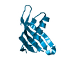
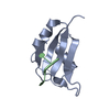
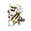
 Streptomyces verticillus (bacteria)
 X-RAY DIFFRACTION /
  Authors
 Citation
 Structure visualization
 Downloads & links
 Other downloads


 PDBj
PDBj
 Assembly

 Mass: 62.068 Da / Num. of mol.: 1 / Source method: obtained synthetically / Formula: C2H6O2
 Mass: 120.147 Da / Num. of mol.: 1 / Source method: obtained synthetically / Formula: C5H12O3 / Comment: precipitant*YM
 Mass: 18.015 Da / Num. of mol.: 31 / Source method: isolated from a natural source / Formula: H2O
 Sample preparation
 / Beamline: 19-BM / Wavelength: 0.97931 Å
 Processing