 Movie
Movie Controller

[English] 日本語
 Yorodumi
- PDB-4zyc: Discovery of dihydroisoquinolinone derivatives as novel inhibitor... -
+ Open data

- Basic information
| Entry | Database: PDB / ID: 4zyc | ||||||
|---|---|---|---|---|---|---|---|
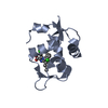
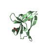
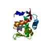
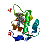
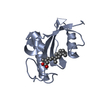
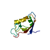
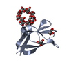
| Title | Discovery of dihydroisoquinolinone derivatives as novel inhibitors of the p53-MDM2 interaction with a distinct binding mode: Hdm2 (MDM2) complexed with cpd5 | ||||||
 Components Components | E3 ubiquitin-protein ligase Mdm2 | ||||||
 Keywords Keywords | LIGASE / PPI WITH P53 / INHIBITOR COMPLEX | ||||||
| Function / homology |  Function and homology information Function and homology informationcellular response to vitamin B1 / response to formaldehyde / response to water-immersion restraint stress / response to ether / traversing start control point of mitotic cell cycle / atrial septum development / fibroblast activation / regulation of protein catabolic process at postsynapse, modulating synaptic transmission / Trafficking of AMPA receptors / ventricular septum development ...cellular response to vitamin B1 / response to formaldehyde / response to water-immersion restraint stress / response to ether / traversing start control point of mitotic cell cycle / atrial septum development / fibroblast activation / regulation of protein catabolic process at postsynapse, modulating synaptic transmission / Trafficking of AMPA receptors / ventricular septum development / negative regulation of intrinsic apoptotic signaling pathway by p53 class mediator / receptor serine/threonine kinase binding / negative regulation of protein processing / response to steroid hormone / SUMO transferase activity / peroxisome proliferator activated receptor binding / positive regulation of vascular associated smooth muscle cell migration / atrioventricular valve morphogenesis / AKT phosphorylates targets in the cytosol / response to iron ion / NEDD8 ligase activity / endocardial cushion morphogenesis / regulation of protein catabolic process / positive regulation of muscle cell differentiation / cellular response to peptide hormone stimulus / blood vessel development / cellular response to alkaloid / cardiac septum morphogenesis / regulation of postsynaptic neurotransmitter receptor internalization / SUMOylation of ubiquitinylation proteins / cellular response to antibiotic / Constitutive Signaling by AKT1 E17K in Cancer / ligase activity / negative regulation of DNA damage response, signal transduction by p53 class mediator / SUMOylation of transcription factors / negative regulation of signal transduction by p53 class mediator / blood vessel remodeling / cellular response to UV-C / cellular response to estrogen stimulus / protein sumoylation / response to magnesium ion / centriolar satellite / protein localization to nucleus / ribonucleoprotein complex binding / positive regulation of vascular associated smooth muscle cell proliferation / protein autoubiquitination / transcription repressor complex / positive regulation of mitotic cell cycle / NPAS4 regulates expression of target genes / regulation of heart rate / positive regulation of protein export from nucleus / : / DNA damage response, signal transduction by p53 class mediator / response to cocaine / sperm end piece / sperm principal piece / establishment of protein localization / ubiquitin binding / Stabilization of p53 / protein destabilization / cellular response to gamma radiation / Regulation of RUNX3 expression and activity / RING-type E3 ubiquitin transferase / cellular response to growth factor stimulus / Oncogene Induced Senescence / Regulation of TP53 Activity through Methylation / Degradation of CDH1 / response to toxic substance / cellular response to hydrogen peroxide / disordered domain specific binding / protein polyubiquitination / ubiquitin-protein transferase activity / p53 binding / endocytic vesicle membrane / Signaling by ALK fusions and activated point mutants / Regulation of TP53 Degradation / ubiquitin protein ligase activity / negative regulation of neuron projection development / positive regulation of proteasomal ubiquitin-dependent protein catabolic process / sperm midpiece / protein-containing complex assembly / 5S rRNA binding / cellular response to hypoxia / Oxidative Stress Induced Senescence / Regulation of TP53 Activity through Phosphorylation / amyloid fibril formation / ubiquitin-dependent protein catabolic process / proteasome-mediated ubiquitin-dependent protein catabolic process / regulation of cell cycle / postsynaptic density / Ub-specific processing proteases / response to xenobiotic stimulus / protein ubiquitination / protein domain specific binding / response to antibiotic / negative regulation of DNA-templated transcription / positive regulation of gene expression / positive regulation of cell population proliferation / ubiquitin protein ligase binding / apoptotic process Similarity search - Function | ||||||
| Biological species |  Homo sapiens (human) Homo sapiens (human) | ||||||
| Method |  X-RAY DIFFRACTION / SYNCHROTRON / MOLECULAR REPLACEMENT / molecular replacement / Resolution: 1.95 Å X-RAY DIFFRACTION / SYNCHROTRON / MOLECULAR REPLACEMENT / molecular replacement / Resolution: 1.95 Å | ||||||
 Authors Authors | Kallen, J. | ||||||
 Citation Citation | Journal: Bioorg.Med.Chem.Lett. / Year: 2015 Title: Discovery of dihydroisoquinolinone derivatives as novel inhibitors of the p53-MDM2 interaction with a distinct binding mode. Authors: Gessier, F. / Kallen, J. / Jacoby, E. / Chene, P. / Stachyra-Valat, T. / Ruetz, S. / Jeay, S. / Holzer, P. / Masuya, K. / Furet, P. | ||||||
| History |
|
- Structure visualization
| Structure viewer | Molecule: MolmilJmol/JSmol |
|---|
- Downloads & links
-Download
| PDBx/mmCIF format | 4zyc.cif.gz | 70.7 KB | Display | PDBx/mmCIF format |
|---|---|---|---|---|
| PDB format | pdb4zyc.ent.gz | 52.5 KB | Display | PDB format |
| PDBx/mmJSON format | 4zyc.json.gz | Tree view | PDBx/mmJSON format | |
| Others |  Other downloads Other downloads |
-Validation report
| Arichive directory | https://data.pdbj.org/pub/pdb/validation_reports/zy/4zycftp://data.pdbj.org/pub/pdb/validation_reports/zy/4zyc | HTTPS FTP |
|---|
-Related structure data
| Related structure data | 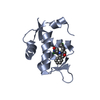 4dijS S: Starting model for refinement |
|---|---|
| Similar structure data |


-Links
 PDBj
PDBj


- Assembly
| Deposited unit | 
| ||||||||
|---|---|---|---|---|---|---|---|---|---|
| 1 | 
| ||||||||
| 2 | 
| ||||||||
| 3 | 
| ||||||||
| Unit cell |
|
-Components
| #1: Protein | Mass: 11172.008 Da / Num. of mol.: 3 Fragment: N-TERMINAL DOMAIN, P53-BINDING DOMAIN, UNP residues 17-111 Mutation: L33E Source method: isolated from a genetically manipulated source Source: (gene. exp.) Homo sapiens (human) / Gene: MDM2 / Production host:  References: UniProt: Q00987, Ligases; Forming carbon-nitrogen bonds; Acid-amino-acid ligases (peptide synthases) #2: Chemical |   Mass: 534.006 Da / Num. of mol.: 3 / Source method: obtained synthetically / Formula: C28H28ClN5O4 Mass: 534.006 Da / Num. of mol.: 3 / Source method: obtained synthetically / Formula: C28H28ClN5O4#3: Chemical | ChemComp-SO4 / |   Mass: 96.063 Da / Num. of mol.: 1 / Source method: obtained synthetically / Formula: SO4 Mass: 96.063 Da / Num. of mol.: 1 / Source method: obtained synthetically / Formula: SO4#4: Water | ChemComp-HOH / |  Mass: 18.015 Da / Num. of mol.: 104 / Source method: isolated from a natural source / Formula: H2O Mass: 18.015 Da / Num. of mol.: 104 / Source method: isolated from a natural source / Formula: H2O |
|---|
-Experimental details
-Experiment
| Experiment | Method: X-RAY DIFFRACTION / Number of used crystals: 1 |
|---|
- Sample preparation
| Crystal | Density Matthews: 0 Å3/Da / Density % sol: 0 % |
|---|---|
| Crystal grow | Temperature: 298 K / Method: vapor diffusion, hanging drop / pH: 8 Details: reservoir: 2.2M ammonium sulphate, 0.2M KNa tartrate, protein: 10mg/ml Hdm2 in 50mM TRIS pH 8.0, 200mM NaCl, 1mM TCEP, 10% glycerol, drop: 0.2ul reservoir + 0.2ul protein PH range: 8 |
-Data collection
| Diffraction | Mean temperature: 100 K |
|---|---|
| Diffraction source | Source: SYNCHROTRON / Site: SLS  / Beamline: X10SA / Wavelength: 0.9794 Å / Beamline: X10SA / Wavelength: 0.9794 Å |
| Detector | Type: MARRESEARCH / Detector: CCD / Date: Nov 7, 2008 |
| Radiation | Monochromator: SI 111 CHANNEL / Protocol: SINGLE WAVELENGTH / Monochromatic (M) / Laue (L): M / Scattering type: x-ray |
| Radiation wavelength | Wavelength: 0.9794 Å / Relative weight: 1 |
| Reflection | Resolution: 1.95→20 Å / Num. obs: 21169 / % possible obs: 99.8 % / Redundancy: 5.3 % / Rmerge(I) obs: 0.056 / Χ2: 0.979 / Net I/av σ(I): 31.07 / Net I/σ(I): 31.07 / Num. measured all: 112750 |
| Reflection shell | Resolution: 1.95→2.02 Å / Redundancy: 5.3 % / Rmerge(I) obs: 0.25 / Mean I/σ(I) obs: 4.21 / Num. unique all: 2147 / Χ2: 1.789 / Rejects: 0 / % possible all: 100 |
-Phasing
| Phasing | Method: molecular replacement |
|---|
- Processing
| Software |
| |||||||||||||||||||||||||||||||||||||||||||||
|---|---|---|---|---|---|---|---|---|---|---|---|---|---|---|---|---|---|---|---|---|---|---|---|---|---|---|---|---|---|---|---|---|---|---|---|---|---|---|---|---|---|---|---|---|---|---|
| Refinement | Method to determine structure: MOLECULAR REPLACEMENT Starting model: 4DIJ Resolution: 1.95→20 Å / Cor.coef. Fo:Fc: 0.937 / Cor.coef. Fo:Fc free: 0.921 / WRfactor Rfree: 0.2718 / WRfactor Rwork: 0.2326 / FOM work R set: 0.8309 / SU B: 3.718 / SU ML: 0.11 / SU R Cruickshank DPI: 0.2044 / SU Rfree: 0.1754 / Cross valid method: THROUGHOUT / σ(F): 0 / ESU R: 0.204 / ESU R Free: 0.175 / Stereochemistry target values: MAXIMUM LIKELIHOOD Details: HYDROGENS HAVE BEEN ADDED IN THE RIDING POSITIONS U VALUES : REFINED INDIVIDUALLY
| |||||||||||||||||||||||||||||||||||||||||||||
| Solvent computation | Ion probe radii: 0.8 Å / Shrinkage radii: 0.8 Å / VDW probe radii: 1.2 Å / Solvent model: MASK | |||||||||||||||||||||||||||||||||||||||||||||
| Displacement parameters | Biso max: 73.82 Å2 / Biso mean: 30.121 Å2 / Biso min: 16.48 Å2
| |||||||||||||||||||||||||||||||||||||||||||||
| Refinement step | Cycle: final / Resolution: 1.95→20 Å
| |||||||||||||||||||||||||||||||||||||||||||||
| Refine LS restraints |
| |||||||||||||||||||||||||||||||||||||||||||||
| LS refinement shell | Resolution: 1.95→2 Å / Total num. of bins used: 20
|
