 Movie
Movie Controller

[English] 日本語
 Yorodumi
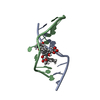
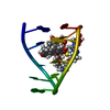
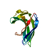
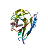
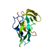
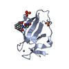
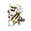
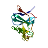
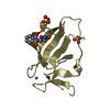
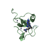
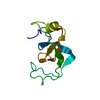
- PDB-2cbm: Crystal structure of the apo-form of a neocarzinostatin mutant ev... -
+ Open data

- Basic information
| Entry | Database: PDB / ID: 2cbm | ||||||
|---|---|---|---|---|---|---|---|
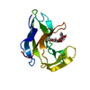
| Title | Crystal structure of the apo-form of a neocarzinostatin mutant evolved to bind testosterone. | ||||||
 Components Components | NEOCARZINOSTATIN | ||||||
 Keywords Keywords | ANTIMICROBIAL / ANTIBIOTIC / DNA-BINDING / PHAGE-DISPLAY | ||||||
| Function / homology |  Function and homology information Function and homology information | ||||||
| Biological species |  STREPTOMYCES CARZINOSTATICUS (bacteria) STREPTOMYCES CARZINOSTATICUS (bacteria) | ||||||
| Method |  X-RAY DIFFRACTION / MOLECULAR REPLACEMENT / Resolution: 2.03 Å X-RAY DIFFRACTION / MOLECULAR REPLACEMENT / Resolution: 2.03 Å | ||||||
 Authors Authors | Drevelle, A. / Graille, M. / Heyd, B. / Sorel, I. / Ulryck, N. / Pecorari, F. / Desmadril, M. / Van Tilbeurgh, H. / Minard, P. | ||||||
 Citation Citation | Journal: J.Mol.Biol. / Year: 2006 Title: Structures of in Vitro Evolved Binding Sites on Neocarzinostatin Scaffold Reveal Unanticipated Evolutionary Pathways. Authors: Drevelle, A. / Graille, M. / Heyd, B. / Sorel, I. / Ulryck, N. / Pecorari, F. / Desmadril, M. / Van Tilbeurgh, H. / Minard, P. #1: Journal: Biochemistry / Year: 2003 Title: In Vitro Evolution of the Binding Specificity of Neocarzinostatin, an Enediyne-Binding Chromoprotein. Authors: Heyd, B. / Pecorari, F. / Collinet, B. / Adjadj, E. / Desmadril, M. / Minard, P. | ||||||
| History |
| ||||||
| Remark 700 | SHEET THE SHEET STRUCTURE OF THIS MOLECULE IS BIFURCATED. IN ORDER TO REPRESENT THIS FEATURE IN ... SHEET THE SHEET STRUCTURE OF THIS MOLECULE IS BIFURCATED. IN ORDER TO REPRESENT THIS FEATURE IN THE SHEET RECORDS BELOW, TWO SHEETS ARE DEFINED. |
- Structure visualization
| Structure viewer | Molecule: MolmilJmol/JSmol |
|---|
- Downloads & links
-Download
| PDBx/mmCIF format | 2cbm.cif.gz | 33.7 KB | Display | PDBx/mmCIF format |
|---|---|---|---|---|
| PDB format | pdb2cbm.ent.gz | 22.7 KB | Display | PDB format |
| PDBx/mmJSON format | 2cbm.json.gz | Tree view | PDBx/mmJSON format | |
| Others |  Other downloads Other downloads |
-Validation report
| Arichive directory | https://data.pdbj.org/pub/pdb/validation_reports/cb/2cbmftp://data.pdbj.org/pub/pdb/validation_reports/cb/2cbm | HTTPS FTP |
|---|
-Related structure data
| Related structure data |  2cboC 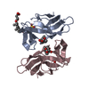 2cbqC 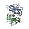 2cbtC 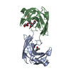 1ncoS S: Starting model for refinement C: citing same article ( |
|---|---|
| Similar structure data |


-Links
 PDBj
PDBj- Assembly
| Deposited unit | 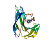
| ||||||||
|---|---|---|---|---|---|---|---|---|---|
| 1 |
| ||||||||
| Unit cell |
| ||||||||
| Components on special symmetry positions |
|
-Components
| #1: Protein | Mass: 11225.162 Da / Num. of mol.: 1 / Fragment: RESIDUES 35-146 / Mutation: YES Source method: isolated from a genetically manipulated source Source: (gene. exp.) STREPTOMYCES CARZINOSTATICUS (bacteria)Production host: |
|---|---|
| #2: Chemical | ChemComp-MES /   Mass: 195.237 Da / Num. of mol.: 1 / Source method: obtained synthetically / Formula: C6H13NO4S / Comment: pH buffer*YM Mass: 195.237 Da / Num. of mol.: 1 / Source method: obtained synthetically / Formula: C6H13NO4S / Comment: pH buffer*YM |
| #3: Water | ChemComp-HOH /  Mass: 18.015 Da / Num. of mol.: 54 / Source method: isolated from a natural source / Formula: H2O Mass: 18.015 Da / Num. of mol.: 54 / Source method: isolated from a natural source / Formula: H2O |
| Compound details | HAS ANTIBIOTIC ACTIVITY FOR GRAM-POSITIVE BACTERIA AND ANTITUMOR ACTIVITY FOR CERTAIN MOUSE TUMORS. ...HAS ANTIBIOTIC |
| Has protein modification | Y |
| Sequence details | D33W,G35A,C37W,W39R,L45W,C47Y,F52N |
-Experimental details
-Experiment
| Experiment | Method: X-RAY DIFFRACTION / Number of used crystals: 1 |
|---|
- Sample preparation
| Crystal | Density Matthews: 2.11 Å3/Da / Density % sol: 41.3 % |
|---|---|
| Crystal grow | Details: 1.8 M (NH4)2SO4; 0.1 M MES PH 6.5 |
-Data collection
| Diffraction | Mean temperature: 100 K |
|---|---|
| Diffraction source | Source: ROTATING ANODE / Type: RIGAKU RU200 / Wavelength: 1.5418 |
| Detector | Type: MARRESEARCH / Detector: IMAGE PLATE |
| Radiation | Protocol: SINGLE WAVELENGTH / Monochromatic (M) / Laue (L): M / Scattering type: x-ray |
| Radiation wavelength | Wavelength: 1.5418 Å / Relative weight: 1 |
| Reflection | Resolution: 2.03→30 Å / Num. obs: 7137 / % possible obs: 98.9 % / Observed criterion σ(I): 1 / Redundancy: 9.38 % / Rmerge(I) obs: 0.09 / Net I/σ(I): 26 |
| Reflection shell | Resolution: 2.03→2.09 Å / Redundancy: 3 % / Rmerge(I) obs: 0.3 / Mean I/σ(I) obs: 8.4 / % possible all: 89.5 |
- Processing
| Software |
| ||||||||||||||||||||||||||||||||||||||||||||||||||||||||||||
|---|---|---|---|---|---|---|---|---|---|---|---|---|---|---|---|---|---|---|---|---|---|---|---|---|---|---|---|---|---|---|---|---|---|---|---|---|---|---|---|---|---|---|---|---|---|---|---|---|---|---|---|---|---|---|---|---|---|---|---|---|---|
| Refinement | Method to determine structure: MOLECULAR REPLACEMENT Starting model: PDB ENTRY 1NCO Resolution: 2.03→30 Å / Data cutoff high absF: 10000 / Cross valid method: THROUGHOUT / σ(F): 0 / Stereochemistry target values: MAXIMUM LIKELIHOOD
| ||||||||||||||||||||||||||||||||||||||||||||||||||||||||||||
| Solvent computation | Bsol: 45.1893 Å2 / ksol: 0.361113 e/Å3 | ||||||||||||||||||||||||||||||||||||||||||||||||||||||||||||
| Displacement parameters |
| ||||||||||||||||||||||||||||||||||||||||||||||||||||||||||||
| Refinement step | Cycle: LAST / Resolution: 2.03→30 Å
| ||||||||||||||||||||||||||||||||||||||||||||||||||||||||||||
| Refine LS restraints |
| ||||||||||||||||||||||||||||||||||||||||||||||||||||||||||||
| Xplor file |
|
