 Movie
Movie Controller

[English] 日本語
 Yorodumi
- PDB-1nco: STRUCTURE OF THE ANTITUMOR PROTEIN-CHROMOPHORE COMPLEX NEOCARZINO... -
+ Open data

- Basic information
| Entry | Database: PDB / ID: 1nco | ||||||
|---|---|---|---|---|---|---|---|
| Title | STRUCTURE OF THE ANTITUMOR PROTEIN-CHROMOPHORE COMPLEX NEOCARZINOSTATIN | ||||||
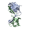
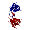
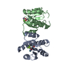
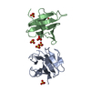
 Components Components | HOLO-NEOCARZINOSTATIN | ||||||
 Keywords Keywords | ANTIBACTERIAL AND ANTITUMOR PROTEIN | ||||||
| Function / homology |  Function and homology information Function and homology information | ||||||
| Biological species |  Streptomyces carzinostaticus (bacteria) Streptomyces carzinostaticus (bacteria) | ||||||
| Method |  X-RAY DIFFRACTION / Resolution: 1.8 Å X-RAY DIFFRACTION / Resolution: 1.8 Å | ||||||
 Authors Authors | Kim, K.-H. / Kwon, B.-M. / Myers, A.G. / Rees, D.C. | ||||||
 Citation Citation | Journal: Science / Year: 1993 Title: Crystal structure of neocarzinostatin, an antitumor protein-chromophore complex. Authors: Kim, K.H. / Kwon, B.M. / Myers, A.G. / Rees, D.C. | ||||||
| History |
|
- Structure visualization
| Structure viewer | Molecule: MolmilJmol/JSmol |
|---|
- Downloads & links
-Download
| PDBx/mmCIF format | 1nco.cif.gz | 55.7 KB | Display | PDBx/mmCIF format |
|---|---|---|---|---|
| PDB format | pdb1nco.ent.gz | 39.8 KB | Display | PDB format |
| PDBx/mmJSON format | 1nco.json.gz | Tree view | PDBx/mmJSON format | |
| Others |  Other downloads Other downloads |
-Validation report
| Arichive directory | https://data.pdbj.org/pub/pdb/validation_reports/nc/1ncoftp://data.pdbj.org/pub/pdb/validation_reports/nc/1nco | HTTPS FTP |
|---|
-Related structure data
| Similar structure data |
|---|

-Links
 PDBj
PDBj- Assembly
| Deposited unit | 
| ||||||||
|---|---|---|---|---|---|---|---|---|---|
| 1 |
| ||||||||
| Unit cell |
| ||||||||
| Atom site foot note | 1: RESIDUES PRO A 9 AND PRO B 9 ARE CIS PROLINES. | ||||||||
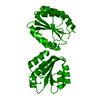
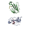
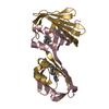
| Details | THE GENERAL NON-CRYSTALLOGRAPHIC TWO-FOLD AXIS BETWEEN TWO MOLECULES IN THE ASYMMETRIC UNIT WAS DETERMINED BY DENSITY CORRELATION STUDIES AND MANUAL MOLECULAR REPLACEMENT WITH MACROMOMYCIN STRUCTURE AS A MODEL. THE MOLECULE PREFIXED WITH "B" IS THE HOLO-NEOCARZINOSTATIN AND THE MOLECULE WITH "A" IS THE APO-NEOCARZINOSTATIN. THE CURRENT MODEL HAS 226 RESIDUES OF MOLECULES A AND B, 1 NEOCARZINOSTATIN-CHROMOPHORE, 1 MPD AND 161 WATER MOLECULES (1773 NON-HYDROGEN ATOMS TOTAL ). |
-Components
| #1: Protein | Mass: 11100.055 Da / Num. of mol.: 2 Source method: isolated from a genetically manipulated source Source: (gene. exp.) Streptomyces carzinostaticus (bacteria)References: UniProt: P0A3R9 #2: Chemical | ChemComp-MRD / ( |   Mass: 118.174 Da / Num. of mol.: 1 / Source method: obtained synthetically / Formula: C6H14O2 / Comment: precipitant*YM Mass: 118.174 Da / Num. of mol.: 1 / Source method: obtained synthetically / Formula: C6H14O2 / Comment: precipitant*YM#3: Chemical | ChemComp-CHR / |   Mass: 659.636 Da / Num. of mol.: 1 / Source method: obtained synthetically / Formula: C35H33NO12 Mass: 659.636 Da / Num. of mol.: 1 / Source method: obtained synthetically / Formula: C35H33NO12#4: Water | ChemComp-HOH / |  Mass: 18.015 Da / Num. of mol.: 161 / Source method: isolated from a natural source / Formula: H2O Mass: 18.015 Da / Num. of mol.: 161 / Source method: isolated from a natural source / Formula: H2OHas protein modification | Y | |
|---|
-Experimental details
-Experiment
| Experiment | Method: X-RAY DIFFRACTION |
|---|
- Sample preparation
| Crystal | Density Matthews: 2.79 Å3/Da / Density % sol: 55.89 % | |||||||||||||||||||||||||
|---|---|---|---|---|---|---|---|---|---|---|---|---|---|---|---|---|---|---|---|---|---|---|---|---|---|---|
| Crystal grow | *PLUS Temperature: 4 ℃ / pH: 5 / Method: vapor diffusion, hanging drop | |||||||||||||||||||||||||
| Components of the solutions | *PLUS
|
-Data collection
| Radiation | Scattering type: x-ray |
|---|---|
| Radiation wavelength | Relative weight: 1 |
| Reflection | *PLUS Highest resolution: 1.7 Å / Num. obs: 23556 / % possible obs: 87.8 % / Num. measured all: 146707 / Rmerge(I) obs: 0.071 |
- Processing
| Software |
| ||||||||||||||||||||||||||||||
|---|---|---|---|---|---|---|---|---|---|---|---|---|---|---|---|---|---|---|---|---|---|---|---|---|---|---|---|---|---|---|---|
| Refinement | Rfactor obs: 0.199 / Highest resolution: 1.8 Å | ||||||||||||||||||||||||||||||
| Refinement step | Cycle: LAST / Highest resolution: 1.8 Å
| ||||||||||||||||||||||||||||||
| Refine LS restraints |
| ||||||||||||||||||||||||||||||
| Refinement | *PLUS Highest resolution: 1.8 Å / Rfactor obs: 0.199 | ||||||||||||||||||||||||||||||
| Solvent computation | *PLUS | ||||||||||||||||||||||||||||||
| Displacement parameters | *PLUS | ||||||||||||||||||||||||||||||
| Refine LS restraints | *PLUS
|
