 Movie
Movie Controller


+ Open data

- Basic information
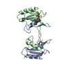
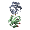
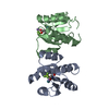
| Entry | Database: PDB / ID: 6z7o | ||||||
|---|---|---|---|---|---|---|---|
| Title | Crystal structure of Thioredoxin T from Drosophila melanogaster | ||||||
 Components Components | Thioredoxin-T | ||||||
 Keywords Keywords | NUCLEAR PROTEIN / Thioredoxin-T / Drosophila melanogaster / Oxidoreductase | ||||||
| Function / homology |  Function and homology information Function and homology informationY chromosome / protein-disulfide reductase activity / protein folding / nucleus Similarity search - Function | ||||||
| Biological species |  | ||||||
| Method |  X-RAY DIFFRACTION / SYNCHROTRON / MOLECULAR REPLACEMENT / Resolution: 2.33 Å X-RAY DIFFRACTION / SYNCHROTRON / MOLECULAR REPLACEMENT / Resolution: 2.33 Å | ||||||
 Authors Authors | Freier, R. / Aragon, E. / Baginski, B. / Pluta, R. / Martin-Malpartida, P. / Torner, C. / Gonzaez, C. / Macias, M. | ||||||
| Funding support |  Spain, 1items Spain, 1items
| ||||||
 Citation Citation | Journal: Iucrj / Year: 2021 Title: Structures of the germline-specific Deadhead and thioredoxin T proteins from Drosophila melanogaster reveal unique features among thioredoxins. Authors: Freier, R. / Aragon, E. / Baginski, B. / Pluta, R. / Martin-Malpartida, P. / Ruiz, L. / Condeminas, M. / Gonzalez, C. / Macias, M.J. #1: Journal: Biorxiv / Year: 2020Title: Structures of the germline-specific Deadhead and Thioredoxin T proteins from Drosophila melanogaster reveal unique features among Thioredoxins Authors: Freier, R. / Aragon, E. / Baginski, B. / Pluta, R. / Martin-Malpartida, P. / Ruiz, L. / Condeminas, M. / Gonzaez, C. / Macias, M. | ||||||
| History |
|
- Structure visualization
| Structure viewer | Molecule: MolmilJmol/JSmol |
|---|
- Downloads & links
-Download
| PDBx/mmCIF format | 6z7o.cif.gz | 61.7 KB | Display | PDBx/mmCIF format |
|---|---|---|---|---|
| PDB format | pdb6z7o.ent.gz | 42.5 KB | Display | PDB format |
| PDBx/mmJSON format | 6z7o.json.gz | Tree view | PDBx/mmJSON format | |
| Others |  Other downloads Other downloads |
-Validation report
| Arichive directory | https://data.pdbj.org/pub/pdb/validation_reports/z7/6z7oftp://data.pdbj.org/pub/pdb/validation_reports/z7/6z7o | HTTPS FTP |
|---|
-Related structure data
| Related structure data |  6zmuC  1xwaS S: Starting model for refinement C: citing same article ( |
|---|---|
| Similar structure data |


-Links
 PDBj
PDBj

- Assembly
| Deposited unit | 
| ||||||||
|---|---|---|---|---|---|---|---|---|---|
| 1 | 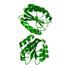
| ||||||||
| Unit cell |
|
-Components
| #1: Protein | Mass: 17515.484 Da / Num. of mol.: 1 Source method: isolated from a genetically manipulated source Details: C-terminus from 107 to 124 (GVYTDEAADVKAVHIDGE) and 129 to 157(LTAESSESDNDNNNVNEVSAHDENAVLEH) not modeled Source: (gene. exp.)  | ||||||
|---|---|---|---|---|---|---|---|
| #2: Chemical |   Mass: 65.409 Da / Num. of mol.: 2 / Source method: obtained synthetically / Formula: Zn Mass: 65.409 Da / Num. of mol.: 2 / Source method: obtained synthetically / Formula: Zn#3: Water | ChemComp-HOH / |  Mass: 18.015 Da / Num. of mol.: 34 / Source method: isolated from a natural source / Formula: H2O Mass: 18.015 Da / Num. of mol.: 34 / Source method: isolated from a natural source / Formula: H2OHas ligand of interest | N | Has protein modification | Y | |
-Experimental details
-Experiment
| Experiment | Method: X-RAY DIFFRACTION / Number of used crystals: 1 |
|---|
- Sample preparation
| Crystal | Density Matthews: 1.89 Å3/Da / Density % sol: 34.86 % |
|---|---|
| Crystal grow | Temperature: 293 K / Method: vapor diffusion / pH: 7.5 / Details: 15.0% w/v PEG 4000, 0.2 M potassium bromide |
-Data collection
| Diffraction | Mean temperature: 100 K / Serial crystal experiment: N | ||||||||||||||||||||||||||||||
|---|---|---|---|---|---|---|---|---|---|---|---|---|---|---|---|---|---|---|---|---|---|---|---|---|---|---|---|---|---|---|---|
| Diffraction source | Source: SYNCHROTRON / Site: ESRF  / Beamline: ID23-2 / Wavelength: 0.872899 Å / Beamline: ID23-2 / Wavelength: 0.872899 Å | ||||||||||||||||||||||||||||||
| Detector | Type: DECTRIS PILATUS3 2M / Detector: PIXEL / Date: Oct 3, 2016 | ||||||||||||||||||||||||||||||
| Radiation | Protocol: SINGLE WAVELENGTH / Monochromatic (M) / Laue (L): M / Scattering type: x-ray | ||||||||||||||||||||||||||||||
| Radiation wavelength | Wavelength: 0.872899 Å / Relative weight: 1 | ||||||||||||||||||||||||||||||
| Reflection | Resolution: 2.32→35.08 Å / Num. obs: 6247 / % possible obs: 99.9 % / Redundancy: 8.1 % / Biso Wilson estimate: 31.19 Å2 / CC1/2: 0.994 / Rmerge(I) obs: 0.219 / Rpim(I) all: 0.079 / Rrim(I) all: 0.233 / Net I/σ(I): 7.4 | ||||||||||||||||||||||||||||||
| Reflection shell | Diffraction-ID: 1
|
- Processing
| Software |
| ||||||||||||||||||||||||||||||
|---|---|---|---|---|---|---|---|---|---|---|---|---|---|---|---|---|---|---|---|---|---|---|---|---|---|---|---|---|---|---|---|
| Refinement | Method to determine structure: MOLECULAR REPLACEMENT Starting model: 1XWA Resolution: 2.33→33.672 Å / SU ML: 0.22 / Cross valid method: THROUGHOUT / σ(F): 1.35 / Phase error: 21.48 / Stereochemistry target values: ML
| ||||||||||||||||||||||||||||||
| Solvent computation | Shrinkage radii: 0.9 Å / VDW probe radii: 1.11 Å / Solvent model: FLAT BULK SOLVENT MODEL | ||||||||||||||||||||||||||||||
| Displacement parameters | Biso max: 114.32 Å2 / Biso mean: 34.4913 Å2 / Biso min: 14.52 Å2 | ||||||||||||||||||||||||||||||
| Refinement step | Cycle: final / Resolution: 2.33→33.672 Å
| ||||||||||||||||||||||||||||||
| Refine LS restraints |
| ||||||||||||||||||||||||||||||
| LS refinement shell | Refine-ID: X-RAY DIFFRACTION / Rfactor Rfree error: 0
|
