 ムービー
ムービー コントローラー


+ データを開く

- 基本情報
基本情報
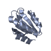
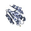
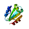
| 登録情報 | データベース: PDB / ID: 1xwa | ||||||
|---|---|---|---|---|---|---|---|
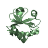
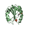
| タイトル | Drospohila thioredoxin, oxidized, P41212 | ||||||
 要素 要素 | thioredoxin | ||||||
 キーワード キーワード | ELECTRON TRANSPORT / Dimerization / Drosophila melanogaster / redox regulation / thioredoxin / x-ray crystal structure | ||||||
| 機能・相同性 |  機能・相同性情報 機能・相同性情報The NLRP3 inflammasome / Protein repair / Detoxification of Reactive Oxygen Species / TP53 Regulates Metabolic Genes / Interconversion of nucleotide di- and triphosphates / Oxidative Stress Induced Senescence / glutathione disulfide oxidoreductase activity / disulfide oxidoreductase activity / protein-disulfide reductase activity / cell redox homeostasis ...The NLRP3 inflammasome / Protein repair / Detoxification of Reactive Oxygen Species / TP53 Regulates Metabolic Genes / Interconversion of nucleotide di- and triphosphates / Oxidative Stress Induced Senescence / glutathione disulfide oxidoreductase activity / disulfide oxidoreductase activity / protein-disulfide reductase activity / cell redox homeostasis / determination of adult lifespan / response to oxidative stress / nucleus / cytosol 類似検索 - 分子機能 | ||||||
| 生物種 |  | ||||||
| 手法 |  X線回折 / シンクロトロン / 分子置換 / 解像度: 2.2 Å X線回折 / シンクロトロン / 分子置換 / 解像度: 2.2 Å | ||||||
 データ登録者 データ登録者 | Wahl, M.C. / Irmler, A. / Hecker, B. / Schirmer, R.H. / Becker, K. | ||||||
 引用 引用 | ジャーナル: J.Mol.Biol. / 年: 2005 タイトル: Comparative structural analysis of oxidized and reduced thioredoxin from Drosophila melanogaster 著者: Wahl, M.C. / Irmler, A. / Hecker, B. / Schirmer, R.H. / Becker, K. | ||||||
| 履歴 |
|
- 構造の表示
構造の表示
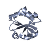
| 構造ビューア | 分子: MolmilJmol/JSmol |
|---|
- ダウンロードとリンク
-ダウンロード
| PDBx/mmCIF形式 | 1xwa.cif.gz | 103.2 KB | 表示 | PDBx/mmCIF形式 |
|---|---|---|---|---|
| PDB形式 | pdb1xwa.ent.gz | 80.3 KB | 表示 | PDB形式 |
| PDBx/mmJSON形式 | 1xwa.json.gz | ツリー表示 | PDBx/mmJSON形式 | |
| その他 |  その他のダウンロード その他のダウンロード |
-検証レポート
| アーカイブディレクトリ | https://data.pdbj.org/pub/pdb/validation_reports/xw/1xwaftp://data.pdbj.org/pub/pdb/validation_reports/xw/1xwa | HTTPS FTP |
|---|
-関連構造データ


-リンク
 PDBj
PDBj

- 集合体
集合体
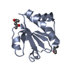
| 登録構造単位 | 
| ||||||||
|---|---|---|---|---|---|---|---|---|---|
| 1 | 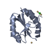
| ||||||||
| 2 | 
| ||||||||
| 3 | 
| ||||||||
| 4 | 
| ||||||||
| 5 | 
| ||||||||
| 6 | 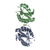
| ||||||||
| 単位格子 |
|
-要素
| #1: タンパク質 | 分子量: 12104.024 Da / 分子数: 4 / 由来タイプ: 組換発現 由来: (組換発現) 遺伝子: TRX-2 / プラスミド: pQE-30 / 生物種 (発現宿主): Escherichia coli / 発現宿主:  #2: 化合物 |   分子量: 112.411 Da / 分子数: 3 / 由来タイプ: 合成 / 式: Cd 分子量: 112.411 Da / 分子数: 3 / 由来タイプ: 合成 / 式: Cd#3: 化合物 | ChemComp-CL / |   分子量: 35.453 Da / 分子数: 1 / 由来タイプ: 合成 / 式: Cl 分子量: 35.453 Da / 分子数: 1 / 由来タイプ: 合成 / 式: Cl#4: 水 | ChemComp-HOH / |  分子量: 18.015 Da / 分子数: 418 / 由来タイプ: 天然 / 式: H2O 分子量: 18.015 Da / 分子数: 418 / 由来タイプ: 天然 / 式: H2OHas protein modification | Y | |
|---|
-実験情報
-実験
| 実験 | 手法: X線回折 / 使用した結晶の数: 1 |
|---|
- 試料調製
試料調製
| 結晶 | マシュー密度: 2.2 Å3/Da / 溶媒含有率: 43.2 % |
|---|---|
| 結晶化 | 温度: 298 K / 手法: 蒸気拡散法 / pH: 4.6 詳細: CdCl2, PEG400, pH 4.6, VAPOR DIFFUSION, temperature 298K |
-データ収集
| 回折 | 平均測定温度: 100 K |
|---|---|
| 放射光源 | 由来: シンクロトロン / サイト: MPG/DESY, HAMBURG  / ビームライン: BW6 / 波長: 1.05 Å / ビームライン: BW6 / 波長: 1.05 Å |
| 検出器 | タイプ: MARRESEARCH / 検出器: CCD / 詳細: mirrors |
| 放射 | プロトコル: SINGLE WAVELENGTH / 単色(M)・ラウエ(L): M / 散乱光タイプ: x-ray |
| 放射波長 | 波長: 1.05 Å / 相対比: 1 |
| 反射 | 解像度: 2.1→30 Å / Num. obs: 37273 / % possible obs: 94.1 % / Observed criterion σ(F): 0 / Observed criterion σ(I): 0 / 冗長度: 5.3 % / Biso Wilson estimate: 35 Å2 / Rsym value: 0.038 / Net I/σ(I): 21.1 |
| 反射 シェル | 解像度: 2.1→2.2 Å / % possible all: 88.1 |
- 解析
解析
| ソフトウェア |
| ||||||||||||||||||||
|---|---|---|---|---|---|---|---|---|---|---|---|---|---|---|---|---|---|---|---|---|---|
| 精密化 | 構造決定の手法: 分子置換 開始モデル: PDB Entry 1AUC 解像度: 2.2→20 Å / 交差検証法: THROUGHOUT / σ(F): 0 / σ(I): 0 / 立体化学のターゲット値: Engh & Huber
| ||||||||||||||||||||
| 精密化ステップ | サイクル: LAST / 解像度: 2.2→20 Å
|
