 Movie
Movie Controller

[English] 日本語
 Yorodumi
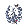
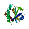
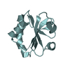
- PDB-2h6x: Crystal Structure of Thioredoxin Wild Type in Hexagonal (p61) Spa... -
+ Open data

- Basic information
| Entry | Database: PDB / ID: 2h6x | ||||||
|---|---|---|---|---|---|---|---|
| Title | Crystal Structure of Thioredoxin Wild Type in Hexagonal (p61) Space Group | ||||||
 Components Components | Thioredoxin | ||||||
 Keywords Keywords | ELECTRON TRANSPORT / Alpha Beta | ||||||
| Function / homology |  Function and homology information Function and homology informationDNA polymerase processivity factor activity / protein-disulfide reductase activity / cell redox homeostasis / cytosol / cytoplasm Similarity search - Function | ||||||
| Biological species |  | ||||||
| Method |  X-RAY DIFFRACTION / MOLECULAR REPLACEMENT / Resolution: 2.6 Å X-RAY DIFFRACTION / MOLECULAR REPLACEMENT / Resolution: 2.6 Å | ||||||
 Authors Authors | Gavira, J.A. / Godoy-Ruiz, R. / Ibarra-Molero, B. / Sanchez-Ruiz, J.M. | ||||||
 Citation Citation | Journal: To be Published Title: Crystal Structure of Thioredoxin Wild Type in Hexagonal (p61) Space Group Authors: Godoy-Ruiz, R. / Gavira, J.A. / Ibarra-Molero, B. / Sanchez-Ruiz, J.M. #1: Journal: J.Mol.Biol. / Year: 2004Title: Relation between protein stability, evolution and structure, as probed by carboxylic acid mutations Authors: Godoy-Ruiz, R. / Perez-Jimenez, R. / Ibarra-Molero, B. / Sanchez-Ruiz, J.M. | ||||||
| History |
|
- Structure visualization
| Structure viewer | Molecule: MolmilJmol/JSmol |
|---|
- Downloads & links
-Download
| PDBx/mmCIF format | 2h6x.cif.gz | 56.4 KB | Display | PDBx/mmCIF format |
|---|---|---|---|---|
| PDB format | pdb2h6x.ent.gz | 40.7 KB | Display | PDB format |
| PDBx/mmJSON format | 2h6x.json.gz | Tree view | PDBx/mmJSON format | |
| Others |  Other downloads Other downloads |
-Validation report
| Arichive directory | https://data.pdbj.org/pub/pdb/validation_reports/h6/2h6xftp://data.pdbj.org/pub/pdb/validation_reports/h6/2h6x | HTTPS FTP |
|---|
-Related structure data
| Related structure data | 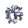 2trxS S: Starting model for refinement |
|---|---|
| Similar structure data |


-Links
 PDBj
PDBj

- Assembly
| Deposited unit | 
| ||||||||
|---|---|---|---|---|---|---|---|---|---|
| 1 | 
| ||||||||
| 2 | 
| ||||||||
| Unit cell |
|
-Components
| #1: Protein | Mass: 11687.388 Da / Num. of mol.: 2 Source method: isolated from a genetically manipulated source Source: (gene. exp.) #2: Chemical |   Mass: 118.174 Da / Num. of mol.: 2 / Source method: obtained synthetically / Formula: C6H14O2 / Comment: precipitant*YM Mass: 118.174 Da / Num. of mol.: 2 / Source method: obtained synthetically / Formula: C6H14O2 / Comment: precipitant*YM#3: Water | ChemComp-HOH / |  Mass: 18.015 Da / Num. of mol.: 83 / Source method: isolated from a natural source / Formula: H2O Mass: 18.015 Da / Num. of mol.: 83 / Source method: isolated from a natural source / Formula: H2OHas protein modification | Y | |
|---|
-Experimental details
-Experiment
| Experiment | Method: X-RAY DIFFRACTION / Number of used crystals: 1 |
|---|
- Sample preparation
| Crystal | Density Matthews: 2.79 Å3/Da / Density % sol: 55.95 % |
|---|---|
| Crystal grow | Temperature: 277 K / Method: counter-diffusion / pH: 3.5 Details: 60% (v/v) MPD, Ac2Cu 1mM, AcNa 15mM, HEPES 15 mM pH 6.9, pH 3.5, Counterdiffusion, temperature 277K |
-Data collection
| Diffraction | Mean temperature: 100 K |
|---|---|
| Diffraction source | Source: ROTATING ANODE / Type: OTHER / Wavelength: 1.5418 Å |
| Detector | Type: BRUKER SMART 6000 / Detector: CCD / Date: Mar 28, 2006 / Details: Montel Optics |
| Radiation | Monochromator: Ni Filter / Protocol: SINGLE WAVELENGTH / Monochromatic (M) / Laue (L): M / Scattering type: x-ray |
| Radiation wavelength | Wavelength: 1.5418 Å / Relative weight: 1 |
| Reflection | Resolution: 2.6→51.36 Å / Num. all: 8147 / Num. obs: 8147 / % possible obs: 99.8 % / Redundancy: 7.28 % / Biso Wilson estimate: 51.288 Å2 / Rsym value: 0.0787 / Net I/σ(I): 9.08 |
| Reflection shell | Resolution: 2.6→2.65 Å / Redundancy: 7.28 % / Mean I/σ(I) obs: 2.63 / Num. unique all: 464 / Rsym value: 0.3161 / % possible all: 100 |
- Processing
| Software |
| |||||||||||||||||||||||||||||||||||||||||||||||||||||||||||||||||||||||||||||||||||||||||||||||||||||||||||||||||||||||||||||||||||||||||||||||||||
|---|---|---|---|---|---|---|---|---|---|---|---|---|---|---|---|---|---|---|---|---|---|---|---|---|---|---|---|---|---|---|---|---|---|---|---|---|---|---|---|---|---|---|---|---|---|---|---|---|---|---|---|---|---|---|---|---|---|---|---|---|---|---|---|---|---|---|---|---|---|---|---|---|---|---|---|---|---|---|---|---|---|---|---|---|---|---|---|---|---|---|---|---|---|---|---|---|---|---|---|---|---|---|---|---|---|---|---|---|---|---|---|---|---|---|---|---|---|---|---|---|---|---|---|---|---|---|---|---|---|---|---|---|---|---|---|---|---|---|---|---|---|---|---|---|---|---|---|---|
| Refinement | Method to determine structure: MOLECULAR REPLACEMENT Starting model: PDB entry 2TRX Resolution: 2.6→44.5 Å / Cor.coef. Fo:Fc: 0.935 / Cor.coef. Fo:Fc free: 0.898 / WRfactor Rfree: 0.284 / WRfactor Rwork: 0.219 / SU B: 15.605 / SU ML: 0.325 / Cross valid method: THROUGHOUT / σ(F): 0 / ESU R: 0.95 / ESU R Free: 0.36 / Stereochemistry target values: MAXIMUM LIKELIHOOD / Details: HYDROGENS HAVE BEEN ADDED IN THE RIDING POSITIONS
| |||||||||||||||||||||||||||||||||||||||||||||||||||||||||||||||||||||||||||||||||||||||||||||||||||||||||||||||||||||||||||||||||||||||||||||||||||
| Solvent computation | Ion probe radii: 0.8 Å / Shrinkage radii: 0.8 Å / VDW probe radii: 1.4 Å / Solvent model: MASK | |||||||||||||||||||||||||||||||||||||||||||||||||||||||||||||||||||||||||||||||||||||||||||||||||||||||||||||||||||||||||||||||||||||||||||||||||||
| Displacement parameters | Biso mean: 43.014 Å2
| |||||||||||||||||||||||||||||||||||||||||||||||||||||||||||||||||||||||||||||||||||||||||||||||||||||||||||||||||||||||||||||||||||||||||||||||||||
| Refinement step | Cycle: LAST / Resolution: 2.6→44.5 Å
| |||||||||||||||||||||||||||||||||||||||||||||||||||||||||||||||||||||||||||||||||||||||||||||||||||||||||||||||||||||||||||||||||||||||||||||||||||
| Refine LS restraints |
| |||||||||||||||||||||||||||||||||||||||||||||||||||||||||||||||||||||||||||||||||||||||||||||||||||||||||||||||||||||||||||||||||||||||||||||||||||
| LS refinement shell | Refine-ID: X-RAY DIFFRACTION / Total num. of bins used: 20
|
