 Movie
Movie Controller

[English] 日本語
 Yorodumi
- PDB-1fyr: DIMER FORMATION THROUGH DOMAIN SWAPPING IN THE CRYSTAL STRUCTURE ... -
+ Open data

- Basic information
| Entry | Database: PDB / ID: 1fyr | ||||||
|---|---|---|---|---|---|---|---|
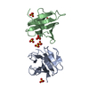
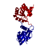
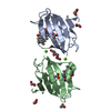
| Title | DIMER FORMATION THROUGH DOMAIN SWAPPING IN THE CRYSTAL STRUCTURE OF THE GRB2-SH2 AC-PYVNV COMPLEX | ||||||
 Components Components |
| ||||||
 Keywords Keywords | HORMONE/GROWTH FACTOR / Grb2 / SH2 domain / phosphopeptide / Met / domain swapping / dimerization / HORMONE-GROWTH FACTOR COMPLEX | ||||||
| Function / homology |  Function and homology information Function and homology information: / : / Regulation of T cell activation by CD28 family / : / Signaling by FGFR3 fusions in cancer / anatomical structure formation involved in morphogenesis / guanyl-nucleotide exchange factor adaptor activity / Grb2-EGFR complex / hepatocyte growth factor receptor activity / Drug-mediated inhibition of MET activation ...: / : / Regulation of T cell activation by CD28 family / : / Signaling by FGFR3 fusions in cancer / anatomical structure formation involved in morphogenesis / guanyl-nucleotide exchange factor adaptor activity / Grb2-EGFR complex / hepatocyte growth factor receptor activity / Drug-mediated inhibition of MET activation / MET activates STAT3 / negative regulation of hydrogen peroxide-mediated programmed cell death / MET Receptor Activation / branching involved in labyrinthine layer morphogenesis / MET interacts with TNS proteins / STAT5 Activation / endothelial cell morphogenesis / Co-inhibition by BTLA / neurotrophin TRKA receptor binding / COP9 signalosome / semaphorin receptor activity / Activated NTRK2 signals through PI3K / MET receptor recycling / transmembrane receptor protein tyrosine kinase adaptor activity / Interleukin-15 signaling / pancreas development / negative regulation of natural killer cell mediated cytotoxicity / Signaling by cytosolic FGFR1 fusion mutants / MET activates PTPN11 / hepatocyte growth factor receptor signaling pathway / MET activates RAP1 and RAC1 / vesicle membrane / Sema4D mediated inhibition of cell attachment and migration / CD28 dependent Vav1 pathway / Signaling by LTK / Signal regulatory protein family interactions / positive regulation of endothelial cell chemotaxis / MET activates PI3K/AKT signaling / MET activates PTK2 signaling / Regulation of KIT signaling / epidermal growth factor receptor binding / natural killer cell mediated cytotoxicity / PI-3K cascade:FGFR3 / STAT5 activation downstream of FLT3 ITD mutants / branching morphogenesis of an epithelial tube / PI-3K cascade:FGFR2 / PI-3K cascade:FGFR4 / positive chemotaxis / PI-3K cascade:FGFR1 / GRB2:SOS provides linkage to MAPK signaling for Integrins / endodermal cell differentiation / positive regulation of actin filament polymerization / RHOU GTPase cycle / semaphorin-plexin signaling pathway / RET signaling / regulation of MAPK cascade / Interleukin-3, Interleukin-5 and GM-CSF signaling / negative regulation of epidermal growth factor receptor signaling pathway / PI3K events in ERBB2 signaling / insulin receptor substrate binding / fibroblast growth factor receptor signaling pathway / PI3K Cascade / Role of LAT2/NTAL/LAB on calcium mobilization / Interleukin receptor SHC signaling / SOS-mediated signalling / Signal attenuation / Activated NTRK3 signals through RAS / Activated NTRK2 signals through RAS / GAB1 signalosome / RHO GTPases Activate WASPs and WAVEs / SHC1 events in ERBB4 signaling / Signalling to RAS / Schwann cell development / Regulation of MITF-M-dependent genes involved in cell cycle and proliferation / SHC-related events triggered by IGF1R / Activated NTRK2 signals through FRS2 and FRS3 / positive regulation of Rac protein signal transduction / SHC-mediated cascade:FGFR3 / MET activates RAS signaling / ephrin receptor binding / SHC-mediated cascade:FGFR2 / SHC-mediated cascade:FGFR4 / Signaling by PDGFRA transmembrane, juxtamembrane and kinase domain mutants / Signaling by PDGFRA extracellular domain mutants / Erythropoietin activates RAS / SHC-mediated cascade:FGFR1 / Signaling by FGFR4 in disease / Signaling by CSF3 (G-CSF) / MECP2 regulates neuronal receptors and channels / FRS-mediated FGFR3 signaling / Signaling by FLT3 ITD and TKD mutants / phosphotyrosine residue binding / signal transduction in response to DNA damage / FRS-mediated FGFR2 signaling / FRS-mediated FGFR4 signaling / FRS-mediated FGFR1 signaling / Signaling by FGFR3 in disease / Tie2 Signaling / myelination / Signaling by FGFR2 in disease Similarity search - Function | ||||||
| Biological species |  Homo sapiens (human) Homo sapiens (human) | ||||||
| Method |  X-RAY DIFFRACTION / SYNCHROTRON / MOLECULAR REPLACEMENT / Resolution: 2.4 Å X-RAY DIFFRACTION / SYNCHROTRON / MOLECULAR REPLACEMENT / Resolution: 2.4 Å | ||||||
 Authors Authors | Schiering, N. / Casale, E. / Caccia, P. / Giordano, P. / Battistini, C. | ||||||
 Citation Citation | Journal: Biochemistry / Year: 2000 Title: Dimer formation through domain swapping in the crystal structure of the Grb2-SH2-Ac-pYVNV complex. Authors: Schiering, N. / Casale, E. / Caccia, P. / Giordano, P. / Battistini, C. | ||||||
| History |
| ||||||
| Remark 300 | BIOMOLECULE: 1, 2 THIS ENTRY CONTAINS THE CRYSTALLOGRAPHIC ASYMMETRIC UNIT WHICH CONSISTS OF 8 ... BIOMOLECULE: 1, 2 THIS ENTRY CONTAINS THE CRYSTALLOGRAPHIC ASYMMETRIC UNIT WHICH CONSISTS OF 8 CHAIN(S). SEE REMARK 350 FOR INFORMATION ON GENERATING THE BIOLOGICAL MOLECULE(S). Please note it has not been proven that the domain- swapped dimer has biological significance. |
- Structure visualization
| Structure viewer | Molecule: MolmilJmol/JSmol |
|---|
- Downloads & links
-Download
| PDBx/mmCIF format | 1fyr.cif.gz | 99.3 KB | Display | PDBx/mmCIF format |
|---|---|---|---|---|
| PDB format | pdb1fyr.ent.gz | 77 KB | Display | PDB format |
| PDBx/mmJSON format | 1fyr.json.gz | Tree view | PDBx/mmJSON format | |
| Others |  Other downloads Other downloads |
-Validation report
| Arichive directory | https://data.pdbj.org/pub/pdb/validation_reports/fy/1fyrftp://data.pdbj.org/pub/pdb/validation_reports/fy/1fyr | HTTPS FTP |
|---|
-Related structure data
| Related structure data | 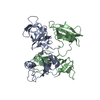 1griS S: Starting model for refinement |
|---|---|
| Similar structure data |


-Links
 PDBj
PDBj


- Assembly
| Deposited unit | 
| ||||||||
|---|---|---|---|---|---|---|---|---|---|
| 1 | 
| ||||||||
| 2 | 
| ||||||||
| 3 | 
| ||||||||
| 4 | 
| ||||||||
| 5 | 
| ||||||||
| 6 | 
| ||||||||
| Unit cell |
| ||||||||
| Details | Dimer 1 is formed from chain I and chain J related by NCS (physiological role not demonstrated) / Dimer 2 is formed from chain K and chain L related by NCS (physiological role not demonstrated) |
-Components
| #1: Protein | Mass: 13281.041 Da / Num. of mol.: 4 / Fragment: SH2 DOMAIN Source method: isolated from a genetically manipulated source Source: (gene. exp.) Homo sapiens (human) / Plasmid: PGEX-2T / Production host:  #2: Protein/peptide | Mass: 599.570 Da / Num. of mol.: 4 / Fragment: RESIDUES 1356-1359 (RESIDUES 0-3 IN COORDINATES) / Source method: obtained synthetically Details: The peptide was chemically synthesized. The sequence occurs naturally in humans. References: UniProt: P08581 #3: Water | ChemComp-HOH / |  Mass: 18.015 Da / Num. of mol.: 182 / Source method: isolated from a natural source / Formula: H2O Mass: 18.015 Da / Num. of mol.: 182 / Source method: isolated from a natural source / Formula: H2OHas protein modification | Y | |
|---|
-Experimental details
-Experiment
| Experiment | Method: X-RAY DIFFRACTION / Number of used crystals: 2 |
|---|
- Sample preparation
| Crystal | Density Matthews: 2.7 Å3/Da / Density % sol: 54 % | ||||||||||||||||||||||||||||||||||||
|---|---|---|---|---|---|---|---|---|---|---|---|---|---|---|---|---|---|---|---|---|---|---|---|---|---|---|---|---|---|---|---|---|---|---|---|---|---|
| Crystal grow | Temperature: 298 K / Method: vapor diffusion, hanging drop / pH: 5.7 Details: 11% PEG 3350, 0.5M NaCL, 0.1M MES/NaOH pH 5.7, VAPOR DIFFUSION, HANGING DROP, temperature 298K | ||||||||||||||||||||||||||||||||||||
| Crystal grow | *PLUS Temperature: 20 ℃ | ||||||||||||||||||||||||||||||||||||
| Components of the solutions | *PLUS
|
-Data collection
| Diffraction | Mean temperature: 100 K |
|---|---|
| Diffraction source | Source: SYNCHROTRON / Site: ELETTRA  / Beamline: 5.2R / Wavelength: 1 / Beamline: 5.2R / Wavelength: 1 |
| Detector | Type: MARRESEARCH / Detector: IMAGE PLATE / Date: Apr 14, 1996 |
| Radiation | Protocol: SINGLE WAVELENGTH / Monochromatic (M) / Laue (L): M / Scattering type: x-ray |
| Radiation wavelength | Wavelength: 1 Å / Relative weight: 1 |
| Reflection | Resolution: 2.4→20 Å / Num. all: 22423 / Num. obs: 22423 / % possible obs: 98.4 % / Observed criterion σ(I): -3 / Redundancy: 6.3 % / Biso Wilson estimate: 32.4 Å2 / Rmerge(I) obs: 0.094 / Net I/σ(I): 16.8 |
| Reflection shell | Resolution: 2.4→2.58 Å / Rmerge(I) obs: 0.252 / % possible all: 97.7 |
| Reflection | *PLUS Num. measured all: 140468 |
| Reflection shell | *PLUS % possible obs: 97.7 % |
- Processing
| Software |
| ||||||||||||||||||||||||||||||||||||
|---|---|---|---|---|---|---|---|---|---|---|---|---|---|---|---|---|---|---|---|---|---|---|---|---|---|---|---|---|---|---|---|---|---|---|---|---|---|
| Refinement | Method to determine structure: MOLECULAR REPLACEMENT Starting model: 1GRI Resolution: 2.4→20 Å / Rfactor Rfree error: 0.008 / Isotropic thermal model: RESTRAINED / Cross valid method: THROUGHOUT / σ(F): 0 / Stereochemistry target values: Engh & Huber
| ||||||||||||||||||||||||||||||||||||
| Solvent computation | Solvent model: mask / Bsol: 30.84 Å2 / ksol: 0.35 e/Å3 | ||||||||||||||||||||||||||||||||||||
| Displacement parameters | Biso mean: 36.8 Å2
| ||||||||||||||||||||||||||||||||||||
| Refine analyze |
| ||||||||||||||||||||||||||||||||||||
| Refinement step | Cycle: LAST / Resolution: 2.4→20 Å
| ||||||||||||||||||||||||||||||||||||
| Refine LS restraints |
| ||||||||||||||||||||||||||||||||||||
| LS refinement shell | Resolution: 2.4→2.55 Å / Rfactor Rfree error: 0.023 / Total num. of bins used: 6
| ||||||||||||||||||||||||||||||||||||
| Xplor file |
| ||||||||||||||||||||||||||||||||||||
| Software | *PLUS Name: CNX / Classification: refinement | ||||||||||||||||||||||||||||||||||||
| Refine LS restraints | *PLUS
|
