 Movie
Movie Controller


+ Open data

- Basic information
| Entry | Database: PDB / ID: 1zdm | ||||||
|---|---|---|---|---|---|---|---|
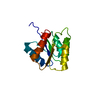
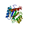
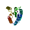
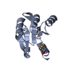
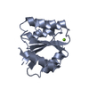
| Title | Crystal Structure of Activated CheY Bound to Xe | ||||||
 Components Components | Chemotaxis protein cheY | ||||||
 Keywords Keywords | SIGNALING PROTEIN / xenon binding / protein cavities / protein conformation assay / activated chey / response regulators / BeF3 | ||||||
| Function / homology |  Function and homology information Function and homology informationbacterial-type flagellum basal body, C ring / bacterial-type flagellum rotor complex / bacterial-type flagellum-dependent swimming motility / aerotaxis / regulation of bacterial-type flagellum-dependent cell motility / thermotaxis / regulation of chemotaxis / internal peptidyl-lysine acetylation / bacterial-type flagellum / phosphorelay response regulator activity ...bacterial-type flagellum basal body, C ring / bacterial-type flagellum rotor complex / bacterial-type flagellum-dependent swimming motility / aerotaxis / regulation of bacterial-type flagellum-dependent cell motility / thermotaxis / regulation of chemotaxis / internal peptidyl-lysine acetylation / bacterial-type flagellum / phosphorelay response regulator activity / protein acetylation / acetyltransferase activity / phosphorelay signal transduction system / chemotaxis / magnesium ion binding / signal transduction / cytosol / cytoplasm Similarity search - Function | ||||||
| Biological species |  | ||||||
| Method |  X-RAY DIFFRACTION / SYNCHROTRON / MOLECULAR REPLACEMENT / Resolution: 2.4 Å X-RAY DIFFRACTION / SYNCHROTRON / MOLECULAR REPLACEMENT / Resolution: 2.4 Å | ||||||
 Authors Authors | Lowery, T.J. / Doucleff, M. / Ruiz, E.J. / Rubin, S.M. / Pines, A. / Wemmer, D.E. | ||||||
 Citation Citation | Journal: Protein Sci. / Year: 2005 Title: Distinguishing multiple chemotaxis Y protein conformations with laser-polarized 129Xe NMR. Authors: Lowery, T.J. / Doucleff, M. / Ruiz, E.J. / Rubin, S.M. / Pines, A. / Wemmer, D.E. #1: Journal: J.Biol.Chem. / Year: 2001Title: Crystal structure of activated CheY. Comparison with other activated receiver domains. Authors: Lee, S.Y. / Cho, H.S. / Pelton, J.G. / Yan, D. / Berry, E.A. / Wemmer, D.E. #2: Journal: J.Mol.Biol. / Year: 2000Title: NMR structure of activated chey Authors: Cho, H.S. / Lee, S.Y. / Yan, D. / Pan, X. / Parkinson, J.S. / Kustu, S. / Wemmer, D.E. / Pelton, J.G. #3: Journal: Nat.Struct.Mol.Biol. / Year: 2001Title: Crystal structure of an activated response regulator bound to its target. Authors: Lee, S.Y. / Cho, H.S. / Pelton, J.G. / Yan, D. / Henderson, R.K. / King, D.S. / Huang, L. / Kustu, S. / Berry, E.A. / Wemmer, D.E. | ||||||
| History |
|
- Structure visualization
| Structure viewer | Molecule: MolmilJmol/JSmol |
|---|
- Downloads & links
-Download
| PDBx/mmCIF format | 1zdm.cif.gz | 66 KB | Display | PDBx/mmCIF format |
|---|---|---|---|---|
| PDB format | pdb1zdm.ent.gz | 48.8 KB | Display | PDB format |
| PDBx/mmJSON format | 1zdm.json.gz | Tree view | PDBx/mmJSON format | |
| Others |  Other downloads Other downloads |
-Validation report
| Arichive directory | https://data.pdbj.org/pub/pdb/validation_reports/zd/1zdmftp://data.pdbj.org/pub/pdb/validation_reports/zd/1zdm | HTTPS FTP |
|---|
-Related structure data
| Related structure data |  1ny5S S: Starting model for refinement |
|---|---|
| Similar structure data |


-Links
 PDBj
PDBj


- Assembly
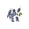
| Deposited unit | 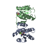
| ||||||||
|---|---|---|---|---|---|---|---|---|---|
| 1 | 
| ||||||||
| 2 | 
| ||||||||
| Unit cell |
|
-Components
| #1: Protein | Mass: 14177.332 Da / Num. of mol.: 2 Source method: isolated from a genetically manipulated source Source: (gene. exp.) #2: Chemical |   Mass: 54.938 Da / Num. of mol.: 2 / Source method: obtained synthetically / Formula: Mn Mass: 54.938 Da / Num. of mol.: 2 / Source method: obtained synthetically / Formula: Mn#3: Chemical |   Mass: 131.293 Da / Num. of mol.: 2 / Source method: obtained synthetically / Formula: Xe Mass: 131.293 Da / Num. of mol.: 2 / Source method: obtained synthetically / Formula: Xe#4: Water | ChemComp-HOH / |  Mass: 18.015 Da / Num. of mol.: 170 / Source method: isolated from a natural source / Formula: H2O Mass: 18.015 Da / Num. of mol.: 170 / Source method: isolated from a natural source / Formula: H2OHas protein modification | Y | |
|---|
-Experimental details
-Experiment
| Experiment | Method: X-RAY DIFFRACTION / Number of used crystals: 1 |
|---|
- Sample preparation
| Crystal | Density Matthews: 4.15 Å3/Da / Density % sol: 70.38 % |
|---|---|
| Crystal grow | Temperature: 298 K / Method: vapor diffusion, hanging drop / pH: 8.4 Details: 1.8 M ammonium sulfate, 5-10% glycerol, 100 mM Tris, pH 8.4, VAPOR DIFFUSION, HANGING DROP, temperature 298K |
-Data collection
| Diffraction | Mean temperature: 100 K |
|---|---|
| Diffraction source | Source: SYNCHROTRON / Site: ALS  / Beamline: 8.3.1 / Wavelength: 1 Å / Beamline: 8.3.1 / Wavelength: 1 Å |
| Detector | Type: ADSC QUANTUM 4 / Detector: CCD / Date: Jul 3, 2003 |
| Radiation | Monochromator: Double crystal / Protocol: SINGLE WAVELENGTH / Monochromatic (M) / Laue (L): M / Scattering type: x-ray |
| Radiation wavelength | Wavelength: 1 Å / Relative weight: 1 |
| Reflection | Resolution: 2.4→80.58 Å / Num. all: 19020 / Num. obs: 19018 / % possible obs: 99.99 % / Observed criterion σ(F): 0 / Observed criterion σ(I): 0 / Rsym value: 0.103 |
| Reflection shell | Resolution: 2.4→2.53 Å / Redundancy: 5.6 % / Mean I/σ(I) obs: 2.2 / Num. unique all: 2801 / Rsym value: 0.439 / % possible all: 99.93 |
- Processing
| Software |
| ||||||||||||||||||||
|---|---|---|---|---|---|---|---|---|---|---|---|---|---|---|---|---|---|---|---|---|---|
| Refinement | Method to determine structure: MOLECULAR REPLACEMENT Starting model: PDB 1NY5 amino acids 1-124 Resolution: 2.4→20 Å / σ(F): 0 / Stereochemistry target values: Engh & Huber
| ||||||||||||||||||||
| Displacement parameters | Biso mean: 52.3 Å2 | ||||||||||||||||||||
| Refine analyze | Luzzati coordinate error obs: 0.34 Å / Luzzati d res low obs: 5 Å / Luzzati sigma a obs: 0.4 Å | ||||||||||||||||||||
| Refinement step | Cycle: LAST / Resolution: 2.4→20 Å
| ||||||||||||||||||||
| Refine LS restraints |
| ||||||||||||||||||||
| LS refinement shell | Resolution: 2.4→2.55 Å / Rfactor Rfree error: 0.021
|
