 Movie
Movie Controller

[English] 日本語
 Yorodumi
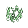
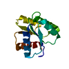
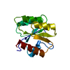
- PDB-2qzj: Crystal structure of a two-component response regulator from Clos... -
+ Open data

- Basic information
| Entry | Database: PDB / ID: 2qzj | ||||||
|---|---|---|---|---|---|---|---|
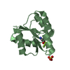
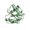
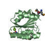
| Title | Crystal structure of a two-component response regulator from Clostridium difficile | ||||||
 Components Components | Two-component response regulator | ||||||
 Keywords Keywords | TRANSCRIPTION / 11017x / PSI-II / Structural Genomics / Protein Structure Initiative / New York SGX Research Center for Structural Genomics / NYSGXRC / DNA-binding / Phosphorylation / Transcription regulation / Two-component regulatory system | ||||||
| Function / homology |  Function and homology information Function and homology informationphosphorelay response regulator activity / protein-DNA complex / transcription cis-regulatory region binding / regulation of DNA-templated transcription / DNA-templated transcription / cytosol Similarity search - Function | ||||||
| Biological species |  Clostridium difficile (bacteria) Clostridium difficile (bacteria) | ||||||
| Method |  X-RAY DIFFRACTION / SYNCHROTRON / SAD / Resolution: 2.89 Å X-RAY DIFFRACTION / SYNCHROTRON / SAD / Resolution: 2.89 Å | ||||||
 Authors Authors | Sugadev, R. / Burley, S.K. / Swaminathan, S. / New York SGX Research Center for Structural Genomics (NYSGXRC) | ||||||
 Citation Citation | Journal: To be Published Title: Crystal structure of a two-component response regulator from Clostridium difficile. Authors: Sugadev, R. / Burley, S.K. / Swaminathan, S. | ||||||
| History |
|
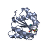
- Structure visualization
| Structure viewer | Molecule: MolmilJmol/JSmol |
|---|
- Downloads & links
-Download
| PDBx/mmCIF format | 2qzj.cif.gz | 152.2 KB | Display | PDBx/mmCIF format |
|---|---|---|---|---|
| PDB format | pdb2qzj.ent.gz | 122.3 KB | Display | PDB format |
| PDBx/mmJSON format | 2qzj.json.gz | Tree view | PDBx/mmJSON format | |
| Others |  Other downloads Other downloads |
-Validation report
| Arichive directory | https://data.pdbj.org/pub/pdb/validation_reports/qz/2qzjftp://data.pdbj.org/pub/pdb/validation_reports/qz/2qzj | HTTPS FTP |
|---|
-Related structure data
| Similar structure data | |
|---|---|
| Other databases |
-Links
 PDBj
PDBj

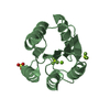
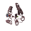
- Assembly
| Deposited unit | 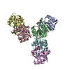
| ||||||||
|---|---|---|---|---|---|---|---|---|---|
| 1 | 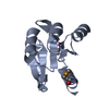
| ||||||||
| 2 | 
| ||||||||
| 3 | 
| ||||||||
| 4 | 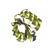
| ||||||||
| 5 | 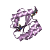
| ||||||||
| 6 | 
| ||||||||
| Unit cell |
|
-Components
| #1: Protein | Mass: 15717.610 Da / Num. of mol.: 6 / Fragment: Residues 2-126 Source method: isolated from a genetically manipulated source Source: (gene. exp.) Clostridium difficile (bacteria) / Strain: 630 / Gene: CD3265 / Plasmid: pSGX3(BC) / Species (production host): Escherichia coli / Production host: #2: Water | ChemComp-HOH / |  Mass: 18.015 Da / Num. of mol.: 78 / Source method: isolated from a natural source / Formula: H2O Mass: 18.015 Da / Num. of mol.: 78 / Source method: isolated from a natural source / Formula: H2OHas protein modification | Y | |
|---|
-Experimental details
-Experiment
| Experiment | Method: X-RAY DIFFRACTION / Number of used crystals: 1 |
|---|
- Sample preparation
| Crystal | Density Matthews: 2.35 Å3/Da / Density % sol: 47.56 % |
|---|---|
| Crystal grow | Temperature: 298 K / Method: vapor diffusion, sitting drop / pH: 5.5 Details: 0.1M Bis-Tris pH 5.5, 25% PEG 3350, VAPOR DIFFUSION, SITTING DROP, temperature 298K |
-Data collection
| Diffraction | Mean temperature: 100 K |
|---|---|
| Diffraction source | Source: SYNCHROTRON / Site: NSLS  / Beamline: X12C / Wavelength: 0.9795 Å / Beamline: X12C / Wavelength: 0.9795 Å |
| Detector | Type: ADSC QUANTUM 210 / Detector: CCD / Date: Aug 11, 2007 / Details: Mirrors |
| Radiation | Monochromator: Si(111) channel / Protocol: SINGLE WAVELENGTH / Monochromatic (M) / Laue (L): M / Scattering type: x-ray |
| Radiation wavelength | Wavelength: 0.9795 Å / Relative weight: 1 |
| Reflection | Resolution: 2.89→50 Å / Num. all: 21182 / Num. obs: 21182 / % possible obs: 99.6 % / Observed criterion σ(F): 0 / Redundancy: 17.7 % / Biso Wilson estimate: 41.1 Å2 / Rmerge(I) obs: 0.069 / Net I/σ(I): 22.8 |
| Reflection shell | Resolution: 2.89→2.99 Å / Redundancy: 13.4 % / Rmerge(I) obs: 0.153 / Mean I/σ(I) obs: 5 / Num. unique all: 2015 / % possible all: 98.5 |
- Processing
| Software |
| |||||||||||||||||||||||||
|---|---|---|---|---|---|---|---|---|---|---|---|---|---|---|---|---|---|---|---|---|---|---|---|---|---|---|
| Refinement | Method to determine structure: SAD / Resolution: 2.89→48.18 Å / Rfactor Rfree error: 0.008 / Data cutoff high absF: 86090.55 / Data cutoff low absF: 0 / Isotropic thermal model: RESTRAINED / Cross valid method: THROUGHOUT / Stereochemistry target values: Engh & Huber Details: Residues listed as missing in Remark 465 are due to lack of electron density. Residues with missing atoms listed in Remark 470 are due to lack of electron density for side chains and modeled as alanines.
| |||||||||||||||||||||||||
| Solvent computation | Solvent model: FLAT MODEL / Bsol: 33.8003 Å2 / ksol: 0.379896 e/Å3 | |||||||||||||||||||||||||
| Displacement parameters | Biso mean: 34.7 Å2
| |||||||||||||||||||||||||
| Refine analyze |
| |||||||||||||||||||||||||
| Refinement step | Cycle: LAST / Resolution: 2.89→48.18 Å
| |||||||||||||||||||||||||
| Refine LS restraints |
| |||||||||||||||||||||||||
| LS refinement shell | Resolution: 2.89→3.07 Å / Rfactor Rfree error: 0.024 / Total num. of bins used: 6
| |||||||||||||||||||||||||
| Xplor file |
|
