 Movie
Movie Controller

[English] 日本語
 Yorodumi
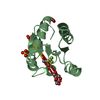
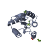
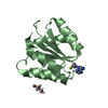
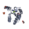
- PDB-3fgz: Crystal Structure of CheY triple mutant F14E, N59M, E89R complexe... -
+ Open data

- Basic information
| Entry | Database: PDB / ID: 3fgz | |||||||||
|---|---|---|---|---|---|---|---|---|---|---|
| Title | Crystal Structure of CheY triple mutant F14E, N59M, E89R complexed with BeF3- and Mn2+ | |||||||||
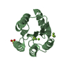
 Components Components | Chemotaxis protein cheY | |||||||||
 Keywords Keywords | SIGNALING PROTEIN / Response Regulator / Receiver Domain / BeF3 / Two-Component Signal Transduction / Chemotaxis / Flagellar rotation / Magnesium / Metal-binding / Phosphoprotein / Two-component regulatory system | |||||||||
| Function / homology |  Function and homology information Function and homology informationbacterial-type flagellum basal body, C ring / bacterial-type flagellum rotor complex / bacterial-type flagellum-dependent swimming motility / aerotaxis / regulation of bacterial-type flagellum-dependent cell motility / regulation of chemotaxis / internal peptidyl-lysine acetylation / thermotaxis / bacterial-type flagellum / phosphorelay response regulator activity ...bacterial-type flagellum basal body, C ring / bacterial-type flagellum rotor complex / bacterial-type flagellum-dependent swimming motility / aerotaxis / regulation of bacterial-type flagellum-dependent cell motility / regulation of chemotaxis / internal peptidyl-lysine acetylation / thermotaxis / bacterial-type flagellum / phosphorelay response regulator activity / acetyltransferase activity / phosphorelay signal transduction system / chemotaxis / magnesium ion binding / signal transduction / cytoplasm / cytosol Similarity search - Function | |||||||||
| Biological species |  | |||||||||
| Method |  X-RAY DIFFRACTION / MOLECULAR REPLACEMENT / Resolution: 2 Å X-RAY DIFFRACTION / MOLECULAR REPLACEMENT / Resolution: 2 Å | |||||||||
 Authors Authors | Wollish, A.C. / Miller, P.J. / Pazy, Y. / Collins, E.J. / Bourret, R.B. | |||||||||
 Citation Citation | Journal: J.Mol.Biol. / Year: 2009 Title: Matching Biochemical Reaction Kinetics to the Timescales of Life: Structural Determinants That Influence the Autodephosphorylation Rate of Response Regulator Proteins. Authors: Pazy, Y. / Wollish, A.C. / Thomas, S.A. / Miller, P.J. / Collins, E.J. / Bourret, R.B. / Silversmith, R.E. | |||||||||
| History |
|
- Structure visualization
| Structure viewer | Molecule: MolmilJmol/JSmol |
|---|
- Downloads & links
-Download
| PDBx/mmCIF format | 3fgz.cif.gz | 67.2 KB | Display | PDBx/mmCIF format |
|---|---|---|---|---|
| PDB format | pdb3fgz.ent.gz | 48.7 KB | Display | PDB format |
| PDBx/mmJSON format | 3fgz.json.gz | Tree view | PDBx/mmJSON format | |
| Others |  Other downloads Other downloads |
-Validation report
| Arichive directory | https://data.pdbj.org/pub/pdb/validation_reports/fg/3fgzftp://data.pdbj.org/pub/pdb/validation_reports/fg/3fgz | HTTPS FTP |
|---|
-Related structure data
| Related structure data | 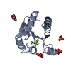 3f7nC 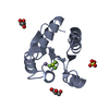 3fftC 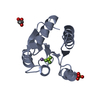 3ffwC 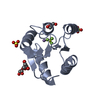 3ffxC  1fqwS S: Starting model for refinement C: citing same article ( |
|---|---|
| Similar structure data |


-Links
 PDBj
PDBj


- Assembly
| Deposited unit | 
| ||||||||
|---|---|---|---|---|---|---|---|---|---|
| 1 | 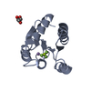
| ||||||||
| 2 | 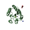
| ||||||||
| Unit cell |
|
-Components
-Protein , 1 types, 2 molecules AB
| #1: Protein | Mass: 14008.249 Da / Num. of mol.: 2 / Mutation: F14E, N59M, E89R Source method: isolated from a genetically manipulated source Source: (gene. exp.) |
|---|
-Non-polymers , 5 types, 176 molecules 


| #2: Chemical |  Mass: 54.938 Da / Num. of mol.: 2 / Source method: obtained synthetically / Formula: Mn Mass: 54.938 Da / Num. of mol.: 2 / Source method: obtained synthetically / Formula: Mn#3: Chemical |  Mass: 66.007 Da / Num. of mol.: 2 / Source method: obtained synthetically / Formula: BeF3 Mass: 66.007 Da / Num. of mol.: 2 / Source method: obtained synthetically / Formula: BeF3#4: Chemical |  Mass: 92.094 Da / Num. of mol.: 2 / Source method: obtained synthetically / Formula: C3H8O3 Mass: 92.094 Da / Num. of mol.: 2 / Source method: obtained synthetically / Formula: C3H8O3#5: Chemical | ChemComp-NH4 / |  Mass: 18.038 Da / Num. of mol.: 1 / Source method: obtained synthetically / Formula: H4N Mass: 18.038 Da / Num. of mol.: 1 / Source method: obtained synthetically / Formula: H4N#6: Water | ChemComp-HOH / | Mass: 18.015 Da / Num. of mol.: 169 / Source method: isolated from a natural source / Formula: H2O |
|---|
-Experimental details
-Experiment
| Experiment | Method: X-RAY DIFFRACTION / Number of used crystals: 1 |
|---|
- Sample preparation
| Crystal | Density Matthews: 4.14 Å3/Da / Density % sol: 70.26 % |
|---|---|
| Crystal grow | Temperature: 298 K / Method: vapor diffusion, hanging drop / pH: 8.4 Details: Tris buffer, Ammonium Sulfate, Glycerol, BeCl2, NaF, MnCl2, pH 8.4, vapor diffusion, hanging drop, temperature 298K |
-Data collection
| Diffraction source | Source: ROTATING ANODE / Type: RIGAKU RUH3R / Wavelength: 1.5418 Å | |||||||||||||||||||||||||||||||||||||||||||||||||||||||||||||||||||||||||||||
|---|---|---|---|---|---|---|---|---|---|---|---|---|---|---|---|---|---|---|---|---|---|---|---|---|---|---|---|---|---|---|---|---|---|---|---|---|---|---|---|---|---|---|---|---|---|---|---|---|---|---|---|---|---|---|---|---|---|---|---|---|---|---|---|---|---|---|---|---|---|---|---|---|---|---|---|---|---|---|
| Detector | Type: RIGAKU RAXIS IV++ / Detector: IMAGE PLATE / Date: Sep 19, 2006 | |||||||||||||||||||||||||||||||||||||||||||||||||||||||||||||||||||||||||||||
| Radiation | Protocol: SINGLE WAVELENGTH / Monochromatic (M) / Laue (L): M / Scattering type: x-ray | |||||||||||||||||||||||||||||||||||||||||||||||||||||||||||||||||||||||||||||
| Radiation wavelength | Wavelength: 1.5418 Å / Relative weight: 1 | |||||||||||||||||||||||||||||||||||||||||||||||||||||||||||||||||||||||||||||
| Reflection | Resolution: 2→80.85 Å / Num. obs: 31410 / % possible obs: 97.2 % / Redundancy: 6.6 % / Rmerge(I) obs: 0.065 / Χ2: 1.086 | |||||||||||||||||||||||||||||||||||||||||||||||||||||||||||||||||||||||||||||
| Reflection shell |
|
- Processing
| Software |
| ||||||||||||||||||||
|---|---|---|---|---|---|---|---|---|---|---|---|---|---|---|---|---|---|---|---|---|---|
| Refinement | Method to determine structure: MOLECULAR REPLACEMENT Starting model: 1FQW Resolution: 2→80.85 Å / Cor.coef. Fo:Fc: 0.943 / Cor.coef. Fo:Fc free: 0.934 / SU ML: 0.078 / ESU R: 0.129 / ESU R Free: 0.122
| ||||||||||||||||||||
| Displacement parameters | Biso mean: 25.411 Å2
| ||||||||||||||||||||
| Refinement step | Cycle: LAST / Resolution: 2→80.85 Å
|
