 Movie
Movie Controller


+ Open data

- Basic information
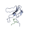
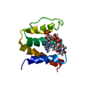
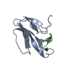
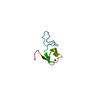
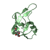
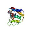
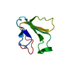
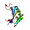
| Entry | Database: PDB / ID: 1hc9 | ||||||
|---|---|---|---|---|---|---|---|
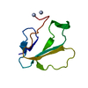
| Title | alpha-bungarotoxin complexed with high affinity peptide | ||||||
 Components Components |
| ||||||
 Keywords Keywords | TOXIN/PEPTIDE / COMPLEX (TOXIN-PEPTIDE) / ACETYLCHOLINE RECEPTOR MIMITOPE / ALPHA-BUNGAROTOXIN / 3- FINGER / PROTEIN-PEPTIDE COMPLEX / TOXIN / TOXIN-PEPTIDE complex | ||||||
| Function / homology |  Function and homology information Function and homology informationacetylcholine receptor inhibitor activity / ion channel regulator activity / toxin activity / extracellular region Similarity search - Function | ||||||
| Biological species |  BUNGARUS MULTICINCTUS (many-banded krait) BUNGARUS MULTICINCTUS (many-banded krait)SYNTHETIC CONSTRUCT (others) | ||||||
| Method |  X-RAY DIFFRACTION / MOLECULAR REPLACEMENT / Resolution: 1.8 Å X-RAY DIFFRACTION / MOLECULAR REPLACEMENT / Resolution: 1.8 Å | ||||||
 Authors Authors | Harel, M. / Kasher, R. / Sussman, J.L. | ||||||
 Citation Citation | Journal: Neuron / Year: 2001 Title: The Binding Site of Acetylcholine Receptor as Visualized in the X-Ray Structure of a Complex between Alpha-Bungarotoxin and a Mimotope Peptide. Authors: Harel, M. / Kasher, R. / Nicolas, A. / Guss, J.M. / Balass, M. / Fridkin, M. / Smit, A.B. / Brejc, K. / Sixma, T.K. / Katchalski-Katzir, E. / Sussman, J.L. / Fuchs, S. | ||||||
| History |
|
- Structure visualization
| Structure viewer | Molecule: MolmilJmol/JSmol |
|---|
- Downloads & links
-Download
| PDBx/mmCIF format | 1hc9.cif.gz | 53.4 KB | Display | PDBx/mmCIF format |
|---|---|---|---|---|
| PDB format | pdb1hc9.ent.gz | 38.8 KB | Display | PDB format |
| PDBx/mmJSON format | 1hc9.json.gz | Tree view | PDBx/mmJSON format | |
| Others |  Other downloads Other downloads |
-Validation report
| Arichive directory | https://data.pdbj.org/pub/pdb/validation_reports/hc/1hc9ftp://data.pdbj.org/pub/pdb/validation_reports/hc/1hc9 | HTTPS FTP |
|---|
-Related structure data
| Related structure data |  1ntnS S: Starting model for refinement |
|---|---|
| Similar structure data |


-Links
 PDBj
PDBj

- Assembly
| Deposited unit | 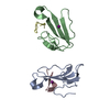
| ||||||||
|---|---|---|---|---|---|---|---|---|---|
| 1 | 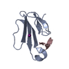
| ||||||||
| 2 | 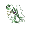
| ||||||||
| Unit cell |
| ||||||||
| Details | COMPLEX OF THE ALPHA-BUNGAROTOXIN AND THE PEPTIDE INHIBITOR |
-Components
| #1: Protein | Mass: 8033.334 Da / Num. of mol.: 1 / Source method: isolated from a natural source / Details: ALPHA-NEUROTOXIN / Source: (natural) BUNGARUS MULTICINCTUS (many-banded krait) / Secretion: VENOM / References: UniProt: P60616 | ||||||||
|---|---|---|---|---|---|---|---|---|---|
| #2: Protein | Mass: 8005.281 Da / Num. of mol.: 1 / Source method: isolated from a natural source / Source: (natural) BUNGARUS MULTICINCTUS (many-banded krait) / Secretion: VENOM / References: UniProt: P60615 | ||||||||
| #3: Protein/peptide | Mass: 1689.843 Da / Num. of mol.: 2 / Source method: obtained synthetically / Details: MIMOTOPE OF THE NICOTINIC ACETYLCHOLINE RECEPTOR / Source: (synth.) SYNTHETIC CONSTRUCT (others) #4: Chemical |   Mass: 126.904 Da / Num. of mol.: 2 / Source method: obtained synthetically / Formula: I Mass: 126.904 Da / Num. of mol.: 2 / Source method: obtained synthetically / Formula: I#5: Water | ChemComp-HOH / |  Mass: 18.015 Da / Num. of mol.: 217 / Source method: isolated from a natural source / Formula: H2O Mass: 18.015 Da / Num. of mol.: 217 / Source method: isolated from a natural source / Formula: H2OCompound details | THE PEPTIDE BINDS ALPHA-BUNGAROTOXIN AND MIMICKS BINDING OF THE TOXIN TO THE NICOTINIC ...THE PEPTIDE BINDS ALPHA-BUNGAROTOX | Has protein modification | Y | |
-Experimental details
-Experiment
| Experiment | Method: X-RAY DIFFRACTION / Number of used crystals: 1 |
|---|
- Sample preparation
| Crystal | Density Matthews: 3.35 Å3/Da / Density % sol: 63 % | ||||||||||||||||||||
|---|---|---|---|---|---|---|---|---|---|---|---|---|---|---|---|---|---|---|---|---|---|
| Crystal grow | pH: 7.5 / Details: 40% PEG 3350, 0.1M PIPES BUFFER PH 7.5 | ||||||||||||||||||||
| Crystal grow | *PLUS Temperature: 19 ℃ / Method: vapor diffusion, hanging drop | ||||||||||||||||||||
| Components of the solutions | *PLUS
|
-Data collection
| Diffraction | Mean temperature: 100 K |
|---|---|
| Diffraction source | Source: ROTATING ANODE / Wavelength: 1.5418 |
| Detector | Type: RIGAKU / Date: Jul 15, 2000 / Details: MIRRORS |
| Radiation | Protocol: SINGLE WAVELENGTH / Monochromatic (M) / Laue (L): M / Scattering type: x-ray |
| Radiation wavelength | Wavelength: 1.5418 Å / Relative weight: 1 |
| Reflection | Resolution: 1.8→23.5 Å / Num. obs: 19144 / % possible obs: 99 % / Redundancy: 6.1 % / Biso Wilson estimate: 16.7 Å2 / Rmerge(I) obs: 0.056 / Rsym value: 0.081 / Net I/σ(I): 14 |
| Reflection shell | Resolution: 1.8→1.86 Å / Redundancy: 5.6 % / Rmerge(I) obs: 0.555 / Mean I/σ(I) obs: 2.3 / Rsym value: 0.594 / % possible all: 99.1 |
| Reflection | *PLUS Num. obs: 22265 / % possible obs: 99 % / Rmerge(I) obs: 0.081 |
| Reflection shell | *PLUS % possible obs: 99.1 % / Rmerge(I) obs: 0.594 |
- Processing
| Software |
| ||||||||||||||||||||||||||||||||||||||||||||||||||||||||||||||||||||||||||||||||
|---|---|---|---|---|---|---|---|---|---|---|---|---|---|---|---|---|---|---|---|---|---|---|---|---|---|---|---|---|---|---|---|---|---|---|---|---|---|---|---|---|---|---|---|---|---|---|---|---|---|---|---|---|---|---|---|---|---|---|---|---|---|---|---|---|---|---|---|---|---|---|---|---|---|---|---|---|---|---|---|---|---|
| Refinement | Method to determine structure: MOLECULAR REPLACEMENT Starting model: PDB ENTRY 1NTN 1-66 RESIDUES Resolution: 1.8→23.47 Å / Rfactor Rfree error: 0.005 / Isotropic thermal model: RESTRAINED / Cross valid method: THROUGHOUT / σ(F): 0 Details: RESIDUES WITH ALTERNATE CONFORMATIONS: A12, A48, A50, A52, A56, A59, B34, B56, B71 THE 2 IODIDE IONS I1 A AND I1 B HAVE OCCUPANCY OF 0.4
| ||||||||||||||||||||||||||||||||||||||||||||||||||||||||||||||||||||||||||||||||
| Solvent computation | Solvent model: FLAT MODEL / Bsol: 70.3 Å2 / ksol: 0.4 e/Å3 | ||||||||||||||||||||||||||||||||||||||||||||||||||||||||||||||||||||||||||||||||
| Displacement parameters | Biso mean: 27.2 Å2
| ||||||||||||||||||||||||||||||||||||||||||||||||||||||||||||||||||||||||||||||||
| Refine analyze |
| ||||||||||||||||||||||||||||||||||||||||||||||||||||||||||||||||||||||||||||||||
| Refinement step | Cycle: LAST / Resolution: 1.8→23.47 Å
| ||||||||||||||||||||||||||||||||||||||||||||||||||||||||||||||||||||||||||||||||
| Refine LS restraints |
| ||||||||||||||||||||||||||||||||||||||||||||||||||||||||||||||||||||||||||||||||
| LS refinement shell | Resolution: 1.8→1.91 Å / Rfactor Rfree error: 0.017 / Total num. of bins used: 6
| ||||||||||||||||||||||||||||||||||||||||||||||||||||||||||||||||||||||||||||||||
| Xplor file |
| ||||||||||||||||||||||||||||||||||||||||||||||||||||||||||||||||||||||||||||||||
| Software | *PLUS Name: CNS / Version: 1 / Classification: refinement | ||||||||||||||||||||||||||||||||||||||||||||||||||||||||||||||||||||||||||||||||
| Refinement | *PLUS Num. reflection obs: 22265 | ||||||||||||||||||||||||||||||||||||||||||||||||||||||||||||||||||||||||||||||||
| Solvent computation | *PLUS | ||||||||||||||||||||||||||||||||||||||||||||||||||||||||||||||||||||||||||||||||
| Displacement parameters | *PLUS | ||||||||||||||||||||||||||||||||||||||||||||||||||||||||||||||||||||||||||||||||
| Refine LS restraints | *PLUS
|
