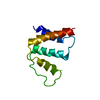
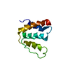
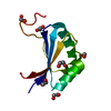
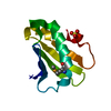
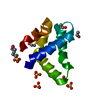
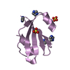
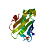
Text: REFINEMENT OF EARLIER PDB DEPOSIT, 1ACA, SEE ENTRY FOR DETAILS. NOE AND DIHEDRAL ANGLE CONSTRAINTS OBTAINED AND MODIFIED FROM THIS ENTRY. RESIDUAL DIPOLAR COUPLINGS ADDED AS ADDITIONAL RESTRAINTS.
-
試料調製
詳細
Solution-ID
内容
溶媒系
1
0.5 mM ACBP, pH 6.5, 0.5 mM palmitoyl-coenzyme A, 5% (3:1) [DMPC:DHPC], 90% H2O, 10% D2O
90% H2O/10% D2O
2
0.5 mM ACBP, pH 6.5, 0.5 mM palmitoyl-coenzyme A, 90% H2O, 10% D2O
90% H2O/10% D2O
試料状態
Conditions-ID
イオン強度
pH
圧 (kPa)
温度 (K)
1
nosaltadded
6.5
ambient
310K
2
nosaltadded
6.5
ambient
298K
-
NMR測定
放射
プロトコル: SINGLE WAVELENGTH / 単色(M)・ラウエ(L): M / 散乱光タイプ: x-ray
放射波長
相対比: 1
NMRスペクトロメーター
タイプ
製造業者
モデル
磁場強度 (MHz)
Spectrometer-ID
Bruker AMX
Bruker
AMX
600
1
Home-built home built
Home-built
homebuilt
600
2
-
解析
NMR software
名称
バージョン
開発者
分類
X-PLOR
modified3.8
Brnger, A. T.
精密化
Pronto
20000515
Kjaeretal.
データ解析
Felix
解析
MNMR
データ解析
精密化
手法: SIMULATED ANNEALING, RESTRAINED MOLECULAR DYNAMICS IN FULL CHARMM FORCE FIELD ソフトェア番号: 1
代表構造
選択基準: lowest energy
NMRアンサンブル
コンフォーマー選択の基準: structures with the lowest energy 計算したコンフォーマーの数: 100 / 登録したコンフォーマーの数: 20
 ムービー
ムービー コントローラー


 データを開く
 基本情報
基本情報 要素
要素 キーワード
キーワード 機能・相同性情報
 データ登録者
 引用
引用 構造の表示
構造の表示 ダウンロードとリンク
 その他のダウンロード
 PDBj
PDBj


 集合体
集合体


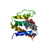
 分子量: 767.534 Da / 分子数: 1 / 由来タイプ: 合成 / 式: C21H36N7O16P3S
 分子量: 256.424 Da / 分子数: 1 / 由来タイプ: 合成 / 式: C16H32O2
 HSQC
HSQC 試料調製
試料調製 解析
解析