 Movie
Movie Controller

[English] 日本語
 Yorodumi
- PDB-2abd: THE THREE-DIMENSIONAL STRUCTURE OF ACYL-COENZYME A BINDING PROTEI... -
+ Open data

- Basic information
| Entry | Database: PDB / ID: 2abd | ||||||
|---|---|---|---|---|---|---|---|
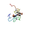
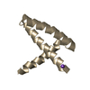
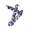
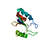
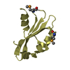
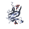
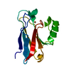
| Title | THE THREE-DIMENSIONAL STRUCTURE OF ACYL-COENZYME A BINDING PROTEIN FROM BOVINE LIVER. STRUCTURAL REFINEMENT USING HETERONUCLEAR MULTIDIMENSIONAL NMR SPECTROSCOPY | ||||||
 Components Components | ACYL-COENZYME A BINDING PROTEIN | ||||||
 Keywords Keywords | ACYL-COENZYME A BINDING PROTEIN | ||||||
| Function / homology |  Function and homology information Function and homology informationMitochondrial Fatty Acid Beta-Oxidation / fatty-acyl-CoA binding / fatty acid metabolic process / Golgi apparatus / endoplasmic reticulum Similarity search - Function | ||||||
| Biological species |  | ||||||
| Method | SOLUTION NMR | ||||||
 Authors Authors | Andersen, K.V. / Poulsen, F.M. | ||||||
 Citation Citation | Journal: J.Biomol.NMR / Year: 1993 Title: The three-dimensional structure of acyl-coenzyme A binding protein from bovine liver: structural refinement using heteronuclear multidimensional NMR spectroscopy. Authors: Andersen, K.V. / Poulsen, F.M. #1: Journal: J.Mol.Biol. / Year: 1992Title: Three-Dimensional Structure in Solution of Acyl-Coenzyme a Binding Protein from Bovine Liver Authors: Andersen, K.V. / Poulsen, F.M. #2: Journal: Biochemistry / Year: 1991Title: The Secondary Structure in Solution of Acyl-Coenzyme a Binding Protein from Bovine Liver Using 1H Nuclear Magnetic Resonance Spectroscopy Authors: Andersen, K.V. / Ludvigsen, S. / Mandrup, S. / Knudsen, J. / Poulsen, F.M. #3: Journal: Biochem.J. / Year: 1987Title: Amino Acid Sequence of Acyl-Coa-Binding Protein from Cow Liver Authors: Mikkelsen, J. / Hojrup, P. / Nielsen, P.R. / Roepstorff, P. / Knudsen, J. | ||||||
| History |
|
- Structure visualization
| Structure viewer | Molecule: MolmilJmol/JSmol |
|---|
- Downloads & links
-Download
| PDBx/mmCIF format | 2abd.cif.gz | 874.5 KB | Display | PDBx/mmCIF format |
|---|---|---|---|---|
| PDB format | pdb2abd.ent.gz | 746.6 KB | Display | PDB format |
| PDBx/mmJSON format | 2abd.json.gz | Tree view | PDBx/mmJSON format | |
| Others |  Other downloads Other downloads |
-Validation report
| Arichive directory | https://data.pdbj.org/pub/pdb/validation_reports/ab/2abdftp://data.pdbj.org/pub/pdb/validation_reports/ab/2abd | HTTPS FTP |
|---|
-Related structure data
| Similar structure data |
|---|


-Links
 PDBj
PDBj
- Assembly
| Deposited unit | 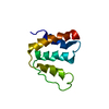
| |||||||||
|---|---|---|---|---|---|---|---|---|---|---|
| 1 |
| |||||||||
| NMR ensembles |
|
-Components
| #1: Protein | Mass: 9931.289 Da / Num. of mol.: 1 Source method: isolated from a genetically manipulated source Source: (gene. exp.)  |
|---|
-Experimental details
-Experiment
| Experiment | Method: SOLUTION NMR |
|---|
- Sample preparation
| Sample conditions | pH: 7 / Temperature: 298 K |
|---|---|
| Crystal grow | *PLUS Method: other / Details: NMR |
- Processing
| Software |
| ||||||||||||
|---|---|---|---|---|---|---|---|---|---|---|---|---|---|
| NMR software |
| ||||||||||||
| NMR ensemble | Conformers submitted total number: 29 |
 X-PLOR