 Movie
Movie Controller

[English] 日本語
 Yorodumi
- PDB-5t9p: Structural analysis reveals the flexible C-terminus of Nop15 unde... -
+ Open data

- Basic information
| Entry | Database: PDB / ID: 5t9p | ||||||
|---|---|---|---|---|---|---|---|
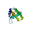
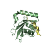
| Title | Structural analysis reveals the flexible C-terminus of Nop15 undergoes rearrangement to recognize a pre-ribosomal RNA folding intermediate | ||||||
 Components Components | Ribosome biogenesis protein 15 | ||||||
 Keywords Keywords | RIBOSOMAL PROTEIN / Nop15 / RRM | ||||||
| Function / homology |  Function and homology information Function and homology informationribosomal large subunit binding / preribosome, large subunit precursor / ribosomal large subunit biogenesis / maturation of LSU-rRNA from tricistronic rRNA transcript (SSU-rRNA, 5.8S rRNA, LSU-rRNA) / rRNA processing / rRNA binding / nucleolus / RNA binding / nucleus / cytoplasm Similarity search - Function | ||||||
| Biological species |  | ||||||
| Method |  X-RAY DIFFRACTION / SYNCHROTRON / MOLECULAR REPLACEMENT / Resolution: 2 Å X-RAY DIFFRACTION / SYNCHROTRON / MOLECULAR REPLACEMENT / Resolution: 2 Å | ||||||
 Authors Authors | Zhang, J. / Gonzalez, E.L. / Hall, M.T.T. | ||||||
| Funding support |  United States, 1items United States, 1items
| ||||||
 Citation Citation | Journal: Nucleic Acids Res / Year: 2017 Title: Structural analysis reveals the flexible C-terminus of Nop15 undergoes rearrangement to recognize a pre-ribosomal RNA folding intermediate. Authors: Jun Zhang / Lauren E Gonzalez / Traci M Tanaka Hall / Abstract: The RNA recognition motif (RRM) is the most abundant RNA-binding domain in eukaryotes, and it plays versatile roles in RNA metabolism. Despite its abundance, diversity of RRM structure and function ...The RNA recognition motif (RRM) is the most abundant RNA-binding domain in eukaryotes, and it plays versatile roles in RNA metabolism. Despite its abundance, diversity of RRM structure and function is generated by variations on a conserved core. Yeast Nop15 is an RRM protein that is essential for large ribosomal subunit biogenesis. We determined a 2.0 Å crystal structure of Nop15 that reveals a C-terminal α-helical region obscures its canonical RNA-binding surface. Small-angle X-ray scattering, NMR and RNA-binding analyses further reveal that the C-terminal residues of Nop15 are highly flexible, but essential for tight RNA binding. Moreover, comparison with a recently reported cryo-electron microscopy structure indicates that dramatic rearrangement of the C-terminal region of Nop15 in the pre-ribosome exposes the RNA-binding surface to recognize the base of its stem-loop target RNA and extends a newly-formed α helix to the distal loop where it forms protein interactions. | ||||||
| History |
|
- Structure visualization
| Structure viewer | Molecule: MolmilJmol/JSmol |
|---|
- Downloads & links
-Download
| PDBx/mmCIF format | 5t9p.cif.gz | 101.9 KB | Display | PDBx/mmCIF format |
|---|---|---|---|---|
| PDB format | pdb5t9p.ent.gz | 78.9 KB | Display | PDB format |
| PDBx/mmJSON format | 5t9p.json.gz | Tree view | PDBx/mmJSON format | |
| Others |  Other downloads Other downloads |
-Validation report
| Arichive directory | https://data.pdbj.org/pub/pdb/validation_reports/t9/5t9pftp://data.pdbj.org/pub/pdb/validation_reports/t9/5t9p | HTTPS FTP |
|---|
-Related structure data
| Related structure data | C: citing same article ( |
|---|---|
| Similar structure data |


-Links
 PDBj
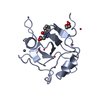
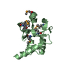
PDBj- Assembly
| Deposited unit | 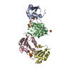
| ||||||||
|---|---|---|---|---|---|---|---|---|---|
| 1 | 
| ||||||||
| 2 | 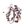
| ||||||||
| 3 | 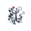
| ||||||||
| 4 | 
| ||||||||
| 5 | 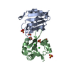
| ||||||||
| 6 | 
| ||||||||
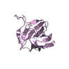
| Unit cell |
|
-Components
| #1: Protein | Mass: 13048.288 Da / Num. of mol.: 4 / Fragment: residues 81-191 Source method: isolated from a genetically manipulated source Source: (gene. exp.) Strain: ATCC 204508 / S288c / Gene: NOP15, YNL110C, N1954 / Production host:  #2: Chemical | ChemComp-SO4 /   Mass: 96.063 Da / Num. of mol.: 11 / Source method: obtained synthetically / Formula: SO4 Mass: 96.063 Da / Num. of mol.: 11 / Source method: obtained synthetically / Formula: SO4#3: Chemical | ChemComp-CL /   Mass: 35.453 Da / Num. of mol.: 7 / Source method: obtained synthetically / Formula: Cl Mass: 35.453 Da / Num. of mol.: 7 / Source method: obtained synthetically / Formula: Cl#4: Water | ChemComp-HOH / |  Mass: 18.015 Da / Num. of mol.: 243 / Source method: isolated from a natural source / Formula: H2O Mass: 18.015 Da / Num. of mol.: 243 / Source method: isolated from a natural source / Formula: H2O |
|---|
-Experimental details
-Experiment
| Experiment | Method: X-RAY DIFFRACTION / Number of used crystals: 1 |
|---|
- Sample preparation
| Crystal | Density Matthews: 2.53 Å3/Da / Density % sol: 51.39 % |
|---|---|
| Crystal grow | Temperature: 295 K / Method: vapor diffusion, hanging drop / pH: 5.25 Details: 18% (w/v) PEG 4000, 0.2 M ammonium sulfate, pH 5.25 and 9% (v/v) Jeffamine 600 |
-Data collection
| Diffraction | Mean temperature: 100 K |
|---|---|
| Diffraction source | Source: SYNCHROTRON / Site: APS / Beamline: 22-ID / Wavelength: 1 Å |
| Detector | Type: MARMOSAIC 225 mm CCD / Detector: CCD / Date: Aug 5, 2012 |
| Radiation | Protocol: SINGLE WAVELENGTH / Monochromatic (M) / Laue (L): M / Scattering type: x-ray |
| Radiation wavelength | Wavelength: 1 Å / Relative weight: 1 |
| Reflection | Resolution: 2→45.06 Å / Num. obs: 33029 / % possible obs: 94.57 % / Redundancy: 3.5 % / Net I/σ(I): 8.53 |
- Processing
| Software |
| |||||||||||||||||||||||||||||||||||||||||||||||||||||||||||||||||||||||||||||||||||||||||||||||||||||||||
|---|---|---|---|---|---|---|---|---|---|---|---|---|---|---|---|---|---|---|---|---|---|---|---|---|---|---|---|---|---|---|---|---|---|---|---|---|---|---|---|---|---|---|---|---|---|---|---|---|---|---|---|---|---|---|---|---|---|---|---|---|---|---|---|---|---|---|---|---|---|---|---|---|---|---|---|---|---|---|---|---|---|---|---|---|---|---|---|---|---|---|---|---|---|---|---|---|---|---|---|---|---|---|---|---|---|---|
| Refinement | Method to determine structure: MOLECULAR REPLACEMENT / Resolution: 2→45 Å / SU ML: 0.25 / Cross valid method: FREE R-VALUE / σ(F): 1.35 / Phase error: 26.02
| |||||||||||||||||||||||||||||||||||||||||||||||||||||||||||||||||||||||||||||||||||||||||||||||||||||||||
| Solvent computation | Shrinkage radii: 0.9 Å / VDW probe radii: 1.11 Å | |||||||||||||||||||||||||||||||||||||||||||||||||||||||||||||||||||||||||||||||||||||||||||||||||||||||||
| Refinement step | Cycle: LAST / Resolution: 2→45 Å
| |||||||||||||||||||||||||||||||||||||||||||||||||||||||||||||||||||||||||||||||||||||||||||||||||||||||||
| Refine LS restraints |
| |||||||||||||||||||||||||||||||||||||||||||||||||||||||||||||||||||||||||||||||||||||||||||||||||||||||||
| LS refinement shell |
|
