 Movie
Movie Controller


+ Open data

- Basic information
| Entry | Database: PDB / ID: 7k3k | |||||||||
|---|---|---|---|---|---|---|---|---|---|---|
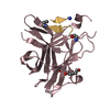
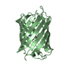
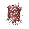
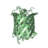
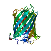
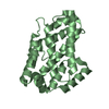
| Title | Crystal structure of dLC8 in complex with Panoramix TQT peptide | |||||||||
 Components Components |
| |||||||||
 Keywords Keywords | MOTOR PROTEIN / Piwi / transposon silencing / heterochromatin formation / piRNA pathway / transcriptional silencing / RNA BINDING PROTEIN | |||||||||
| Function / homology |  Function and homology information Function and homology informationspermatid nucleus elongation / chaeta morphogenesis / Macroautophagy / Aggrephagy / wing disc development / HSP90 chaperone cycle for steroid hormone receptors (SHR) in the presence of ligand / COPI-mediated anterograde transport / positive regulation of neuron remodeling / RNA polymerase II transcription repressor complex / sperm individualization ...spermatid nucleus elongation / chaeta morphogenesis / Macroautophagy / Aggrephagy / wing disc development / HSP90 chaperone cycle for steroid hormone receptors (SHR) in the presence of ligand / COPI-mediated anterograde transport / positive regulation of neuron remodeling / RNA polymerase II transcription repressor complex / sperm individualization / imaginal disc-derived wing morphogenesis / COPI-independent Golgi-to-ER retrograde traffic / piRNA binding / transposable element silencing by piRNA-mediated heterochromatin formation / chaeta development / microtubule anchoring at centrosome / transposable element silencing by heterochromatin formation / Neutrophil degranulation / dynein complex / dynein light intermediate chain binding / cytoplasmic dynein complex / oogenesis / establishment of mitotic spindle orientation / dynein intermediate chain binding / actin filament bundle assembly / transcription repressor complex / centriole / disordered domain specific binding / heterochromatin formation / spermatogenesis / microtubule / structural molecule activity / protein homodimerization activity / protein-containing complex / nucleus / cytosol / cytoplasm Similarity search - Function | |||||||||
| Biological species |  | |||||||||
| Method |  X-RAY DIFFRACTION / SYNCHROTRON / MOLECULAR REPLACEMENT / Resolution: 1.415 Å X-RAY DIFFRACTION / SYNCHROTRON / MOLECULAR REPLACEMENT / Resolution: 1.415 Å | |||||||||
 Authors Authors | Wang, J. / Patel, D.J. | |||||||||
| Funding support |  United States, 2items United States, 2items
| |||||||||
 Citation Citation | Journal: Genes Dev. / Year: 2021 Title: Molecular principles of Piwi-mediated cotranscriptional silencing through the dimeric SFiNX complex. Authors: Schnabl, J. / Wang, J. / Hohmann, U. / Gehre, M. / Batki, J. / Andreev, V.I. / Purkhauser, K. / Fasching, N. / Duchek, P. / Novatchkova, M. / Mechtler, K. / Plaschka, C. / Patel, D.J. / Brennecke, J. | |||||||||
| History |
|
- Structure visualization
| Structure viewer | Molecule: MolmilJmol/JSmol |
|---|
- Downloads & links
-Download
| PDBx/mmCIF format | 7k3k.cif.gz | 39.5 KB | Display | PDBx/mmCIF format |
|---|---|---|---|---|
| PDB format | pdb7k3k.ent.gz | 25 KB | Display | PDB format |
| PDBx/mmJSON format | 7k3k.json.gz | Tree view | PDBx/mmJSON format | |
| Others |  Other downloads Other downloads |
-Validation report
| Arichive directory | https://data.pdbj.org/pub/pdb/validation_reports/k3/7k3kftp://data.pdbj.org/pub/pdb/validation_reports/k3/7k3k | HTTPS FTP |
|---|
-Related structure data
| Related structure data |  7k3jC  7k3lC  3zkeS S: Starting model for refinement C: citing same article ( |
|---|---|
| Similar structure data |


-Links
 PDBj
PDBj

- Assembly
| Deposited unit | 
| ||||||||
|---|---|---|---|---|---|---|---|---|---|
| 1 | 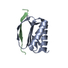
| ||||||||
| Unit cell |
| ||||||||
| Components on special symmetry positions |
|
-Components
| #1: Protein | Mass: 10388.849 Da / Num. of mol.: 1 Source method: isolated from a genetically manipulated source Source: (gene. exp.)  |
|---|---|
| #2: Protein/peptide | Mass: 1641.804 Da / Num. of mol.: 1 / Fragment: residues 445-467 Source method: isolated from a genetically manipulated source Source: (gene. exp.) |
| #3: Water | ChemComp-HOH /  Mass: 18.015 Da / Num. of mol.: 145 / Source method: isolated from a natural source / Formula: H2O Mass: 18.015 Da / Num. of mol.: 145 / Source method: isolated from a natural source / Formula: H2O |
-Experimental details
-Experiment
| Experiment | Method: X-RAY DIFFRACTION / Number of used crystals: 1 |
|---|
- Sample preparation
| Crystal | Density Matthews: 2.53 Å3/Da / Density % sol: 51.44 % |
|---|---|
| Crystal grow | Temperature: 293 K / Method: vapor diffusion, sitting drop Details: 0.1 M NH4Ac, 0.1 M Bis-Tris pH 5.5, 17% (w/v) PEG 10000 |
-Data collection
| Diffraction | Mean temperature: 100 K / Serial crystal experiment: N |
|---|---|
| Diffraction source | Source: SYNCHROTRON / Site: APS / Beamline: 24-ID-E / Wavelength: 0.9792 Å |
| Detector | Type: DECTRIS EIGER X 16M / Detector: PIXEL / Date: Jul 18, 2018 |
| Radiation | Protocol: SINGLE WAVELENGTH / Monochromatic (M) / Laue (L): M / Scattering type: x-ray |
| Radiation wavelength | Wavelength: 0.9792 Å / Relative weight: 1 |
| Reflection | Resolution: 1.415→101.36 Å / Num. obs: 24954 / % possible obs: 99.1 % / Redundancy: 15.6 % / CC1/2: 0.999 / Rmerge(I) obs: 0.069 / Net I/σ(I): 33.6 |
| Reflection shell | Resolution: 1.42→1.44 Å / Num. unique obs: 999 / CC1/2: 0.946 |
- Processing
| Software |
| ||||||||||||||||||||||||||||||||||||||||||||||||||||||||||||||||||||||||||||||||||||||||||
|---|---|---|---|---|---|---|---|---|---|---|---|---|---|---|---|---|---|---|---|---|---|---|---|---|---|---|---|---|---|---|---|---|---|---|---|---|---|---|---|---|---|---|---|---|---|---|---|---|---|---|---|---|---|---|---|---|---|---|---|---|---|---|---|---|---|---|---|---|---|---|---|---|---|---|---|---|---|---|---|---|---|---|---|---|---|---|---|---|---|---|---|
| Refinement | Method to determine structure: MOLECULAR REPLACEMENT Starting model: 3ZKE Resolution: 1.415→38.794 Å / SU ML: 0.12 / Cross valid method: THROUGHOUT / σ(F): 1.35 / Phase error: 18.05 / Stereochemistry target values: ML
| ||||||||||||||||||||||||||||||||||||||||||||||||||||||||||||||||||||||||||||||||||||||||||
| Solvent computation | Shrinkage radii: 0.9 Å / VDW probe radii: 1.11 Å / Solvent model: FLAT BULK SOLVENT MODEL | ||||||||||||||||||||||||||||||||||||||||||||||||||||||||||||||||||||||||||||||||||||||||||
| Displacement parameters | Biso max: 70.88 Å2 / Biso mean: 18.5887 Å2 / Biso min: 8.36 Å2 | ||||||||||||||||||||||||||||||||||||||||||||||||||||||||||||||||||||||||||||||||||||||||||
| Refinement step | Cycle: final / Resolution: 1.415→38.794 Å
| ||||||||||||||||||||||||||||||||||||||||||||||||||||||||||||||||||||||||||||||||||||||||||
| Refine LS restraints |
| ||||||||||||||||||||||||||||||||||||||||||||||||||||||||||||||||||||||||||||||||||||||||||
| LS refinement shell | Refine-ID: X-RAY DIFFRACTION / Rfactor Rfree error: 0
|
