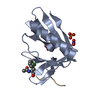
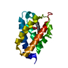
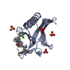
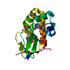
- PDB-2ogb: Crystal structure of the C-terminal domain of mouse Nrdp1 -
+
Open data
ID or keywords:
Loading...
-
Basic information
Entry
Database: PDB / ID: 2ogb
Title
Crystal structure of the C-terminal domain of mouse Nrdp1
Components
RING finger protein 41
Keywords
LIGASE / E3 ubiquitin ligase / Receptor-binding region
Function / homology
Function and homology information
interleukin-3 receptor binding / erythropoietin receptor binding / regulation of lymphocyte differentiation / regulation of myeloid cell differentiation / Downregulation of ERBB2:ERBB3 signaling / endoplasmic reticulum tubular network / regulation of phosphatidylinositol 3-kinase/protein kinase B signal transduction / Antigen processing: Ubiquitination & Proteasome degradation / regulation of reactive oxygen species metabolic process / negative regulation of mitophagy ...interleukin-3 receptor binding / erythropoietin receptor binding / regulation of lymphocyte differentiation / regulation of myeloid cell differentiation / Downregulation of ERBB2:ERBB3 signaling / endoplasmic reticulum tubular network / regulation of phosphatidylinositol 3-kinase/protein kinase B signal transduction / Antigen processing: Ubiquitination & Proteasome degradation / regulation of reactive oxygen species metabolic process / negative regulation of mitophagy / regulation of MAPK cascade / protein autoubiquitination / extrinsic apoptotic signaling pathway / proteasomal protein catabolic process / negative regulation of cell migration / receptor tyrosine kinase binding / RING-type E3 ubiquitin transferase / autophagy / positive regulation of reactive oxygen species metabolic process / small GTPase binding / protein polyubiquitination / ubiquitin-protein transferase activity / positive regulation of protein catabolic process / ubiquitin protein ligase activity / negative regulation of cell population proliferation / protein domain specific binding / perinuclear region of cytoplasm / zinc ion binding / identical protein binding Similarity search - Function
Monochromator: KOHZU double crystal monochromator / Protocol: SINGLE WAVELENGTH / Monochromatic (M) / Laue (L): M / Scattering type: x-ray
Radiation wavelength
Wavelength: 0.9792 Å / Relative weight: 1
Reflection
Redundancy: 4.5 % / Av σ(I) over netI: 26.1 / Number: 89260 / Rmerge(I) obs: 0.092 / Χ2: 1.01 / D res high: 2 Å / D res low: 30 Å / Num. obs: 19750 / % possible obs: 86.9
Diffraction reflection shell
Highest resolution (Å)
Lowest resolution (Å)
% possible obs (%)
ID
Rmerge(I) obs
Chi squared
Redundancy
4.31
30
85.5
1
0.078
0.961
5.3
3.42
4.31
89.5
1
0.074
0.883
5.3
2.99
3.42
91.9
1
0.092
0.948
5.2
2.71
2.99
92.2
1
0.117
0.937
5.2
2.52
2.71
92.3
1
0.143
1.008
5.1
2.37
2.52
93.1
1
0.158
1.079
4.9
2.25
2.37
92.7
1
0.183
1.155
4.2
2.15
2.25
91
1
0.227
1.139
3.7
2.07
2.15
79.7
1
0.275
1.136
2.9
2
2.07
60.9
1
0.306
1.094
2.4
Reflection
Resolution: 1.95→30 Å / Num. obs: 20628 / % possible obs: 85.2 % / Redundancy: 4.3 % / Rmerge(I) obs: 0.092 / Χ2: 0.993 / Net I/σ(I): 25.3
In the structure databanks used in Yorodumi, some data are registered as the other names, "COVID-19 virus" and "2019-nCoV". Here are the details of the virus and the list of structure data.
Jan 31, 2019. EMDB accession codes are about to change! (news from PDBe EMDB page)
EMDB accession codes are about to change! (news from PDBe EMDB page)
The allocation of 4 digits for EMDB accession codes will soon come to an end. Whilst these codes will remain in use, new EMDB accession codes will include an additional digit and will expand incrementally as the available range of codes is exhausted. The current 4-digit format prefixed with “EMD-” (i.e. EMD-XXXX) will advance to a 5-digit format (i.e. EMD-XXXXX), and so on. It is currently estimated that the 4-digit codes will be depleted around Spring 2019, at which point the 5-digit format will come into force.
The EM Navigator/Yorodumi systems omit the EMD- prefix.
Related info.:Q: What is EMD? / ID/Accession-code notation in Yorodumi/EM Navigator
Yorodumi is a browser for structure data from EMDB, PDB, SASBDB, etc.
This page is also the successor to EM Navigator detail page, and also detail information page/front-end page for Omokage search.
The word "yorodu" (or yorozu) is an old Japanese word meaning "ten thousand". "mi" (miru) is to see.
Related info.:EMDB / PDB / SASBDB / Comparison of 3 databanks / Yorodumi Search / Aug 31, 2016. New EM Navigator & Yorodumi / Yorodumi Papers / Jmol/JSmol / Function and homology information / Changes in new EM Navigator and Yorodumi
 Movie
Movie Controller


 Open data
 Basic information
 Components
 Keywords
 Function and homology information
 X-RAY DIFFRACTION /
  Authors
 Citation
 Structure visualization
 Downloads & links
 Other downloads
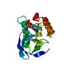


 PDBj
PDBj


 Assembly


 Mass: 58.082 Da / Num. of mol.: 1 / Source method: obtained synthetically / Formula: CNS
 Mass: 92.094 Da / Num. of mol.: 2 / Source method: obtained synthetically / Formula: C3H8O3
 Mass: 18.015 Da / Num. of mol.: 122 / Source method: isolated from a natural source / Formula: H2O
 Sample preparation
 / Beamline: X4A / Wavelength: 0.9792
  Processing