 Movie
Movie Controller


+ Open data

- Basic information
| Entry | Database: PDB / ID: 1kcq | ||||||
|---|---|---|---|---|---|---|---|
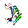
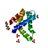
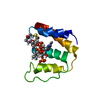
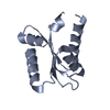
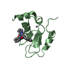
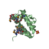
| Title | Human Gelsolin Domain 2 with a Cd2+ bound | ||||||
 Components Components | GELSOLIN | ||||||
 Keywords Keywords | STRUCTURAL PROTEIN / alpha-beta structure / actin-binding protein / familial amyloidosis--Finnish type / cadmium binding / metal binding | ||||||
| Function / homology |  Function and homology information Function and homology informationstriated muscle atrophy / regulation of establishment of T cell polarity / regulation of plasma membrane raft polarization / regulation of receptor clustering / positive regulation of keratinocyte apoptotic process / renal protein absorption / positive regulation of protein processing in phagocytic vesicle / phosphatidylinositol 3-kinase catalytic subunit binding / positive regulation of actin nucleation / actin cap ...striated muscle atrophy / regulation of establishment of T cell polarity / regulation of plasma membrane raft polarization / regulation of receptor clustering / positive regulation of keratinocyte apoptotic process / renal protein absorption / positive regulation of protein processing in phagocytic vesicle / phosphatidylinositol 3-kinase catalytic subunit binding / positive regulation of actin nucleation / actin cap / myosin II binding / host-mediated suppression of symbiont invasion / actin filament severing / barbed-end actin filament capping / cell projection assembly / actin polymerization or depolymerization / actin filament depolymerization / actin filament capping / relaxation of cardiac muscle / Sensory processing of sound by outer hair cells of the cochlea / phagocytosis, engulfment / cardiac muscle cell contraction / hepatocyte apoptotic process / sarcoplasm / cilium assembly / Caspase-mediated cleavage of cytoskeletal proteins / phagocytic vesicle / response to muscle stretch / actin filament polymerization / phosphatidylinositol-4,5-bisphosphate binding / actin filament organization / central nervous system development / protein destabilization / cellular response to type II interferon / actin filament binding / lamellipodium / actin cytoskeleton / actin binding / secretory granule lumen / blood microparticle / amyloid fibril formation / ficolin-1-rich granule lumen / Amyloid fiber formation / focal adhesion / calcium ion binding / Neutrophil degranulation / positive regulation of gene expression / : / extracellular exosome / extracellular region / plasma membrane / cytoplasm / cytosol Similarity search - Function | ||||||
| Biological species |  Homo sapiens (human) Homo sapiens (human) | ||||||
| Method |  X-RAY DIFFRACTION / SYNCHROTRON / MOLECULAR REPLACEMENT / Resolution: 1.65 Å X-RAY DIFFRACTION / SYNCHROTRON / MOLECULAR REPLACEMENT / Resolution: 1.65 Å | ||||||
 Authors Authors | Kazmirski, S.L. / Isaacson, R.L. / An, C. / Buckle, A. / Johnson, C.M. / Daggett, V. / Fersht, A.R. | ||||||
 Citation Citation | Journal: Nat.Struct.Biol. / Year: 2002 Title: Loss of a metal-binding site in gelsolin leads to familial amyloidosis-Finnish type. Authors: Kazmirski, S.L. / Isaacson, R.L. / An, C. / Buckle, A. / Johnson, C.M. / Daggett, V. / Fersht, A.R. | ||||||
| History |
|
- Structure visualization
| Structure viewer | Molecule: MolmilJmol/JSmol |
|---|
- Downloads & links
-Download
| PDBx/mmCIF format | 1kcq.cif.gz | 38.2 KB | Display | PDBx/mmCIF format |
|---|---|---|---|---|
| PDB format | pdb1kcq.ent.gz | 24.5 KB | Display | PDB format |
| PDBx/mmJSON format | 1kcq.json.gz | Tree view | PDBx/mmJSON format | |
| Others |  Other downloads Other downloads |
-Validation report
| Arichive directory | https://data.pdbj.org/pub/pdb/validation_reports/kc/1kcqftp://data.pdbj.org/pub/pdb/validation_reports/kc/1kcq | HTTPS FTP |
|---|
-Related structure data
| Related structure data |  1d0nS S: Starting model for refinement |
|---|---|
| Similar structure data |


-Links
 PDBj
PDBj

- Assembly
| Deposited unit | 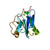
| ||||||||||
|---|---|---|---|---|---|---|---|---|---|---|---|
| 1 |
| ||||||||||
| 2 | 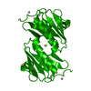
| ||||||||||
| Unit cell |
|
-Components
| #1: Protein | Mass: 11592.965 Da / Num. of mol.: 1 / Fragment: DOMAIN 2 Source method: isolated from a genetically manipulated source Source: (gene. exp.) Homo sapiens (human) / Production host:  | ||||
|---|---|---|---|---|---|
| #2: Chemical | ChemComp-CD /   Mass: 112.411 Da / Num. of mol.: 4 / Source method: obtained synthetically / Formula: Cd Mass: 112.411 Da / Num. of mol.: 4 / Source method: obtained synthetically / Formula: Cd#3: Water | ChemComp-HOH / |  Mass: 18.015 Da / Num. of mol.: 130 / Source method: isolated from a natural source / Formula: H2O Mass: 18.015 Da / Num. of mol.: 130 / Source method: isolated from a natural source / Formula: H2OHas protein modification | Y | |
-Experimental details
-Experiment
| Experiment | Method: X-RAY DIFFRACTION / Number of used crystals: 1 |
|---|
- Sample preparation
| Crystal | Density Matthews: 2.39 Å3/Da / Density % sol: 48.43 % | ||||||||||||||||||||||||||||
|---|---|---|---|---|---|---|---|---|---|---|---|---|---|---|---|---|---|---|---|---|---|---|---|---|---|---|---|---|---|
| Crystal grow | Temperature: 290 K / Method: vapor diffusion, hanging drop / pH: 4.6 Details: PEG 400, cadmium chloride, sodium acetate, pH 4.6, VAPOR DIFFUSION, HANGING DROP at 290K | ||||||||||||||||||||||||||||
| Crystal grow | *PLUS | ||||||||||||||||||||||||||||
| Components of the solutions | *PLUS
|
-Data collection
| Diffraction | Mean temperature: 100 K |
|---|---|
| Diffraction source | Source: SYNCHROTRON / Site: SRS  / Beamline: PX9.5 / Wavelength: 1.2 Å / Beamline: PX9.5 / Wavelength: 1.2 Å |
| Detector | Type: MARRESEARCH / Detector: CCD / Date: Sep 4, 2000 |
| Radiation | Monochromator: Si 111 CHANNEL / Protocol: SINGLE WAVELENGTH / Monochromatic (M) / Laue (L): M / Scattering type: x-ray |
| Radiation wavelength | Wavelength: 1.2 Å / Relative weight: 1 |
| Reflection | Resolution: 1.65→43.033 Å / Num. obs: 14799 / % possible obs: 99 % / Observed criterion σ(I): 1 / Redundancy: 3.3 % / Biso Wilson estimate: 33.8 Å2 / Rmerge(I) obs: 0.075 / Net I/σ(I): 5.2 |
| Reflection shell | Resolution: 1.65→1.74 Å / Redundancy: 2.7 % / Rmerge(I) obs: 0.174 / % possible all: 94.6 |
| Reflection | *PLUS Lowest resolution: 43 Å / Num. obs: 13093 / Num. measured all: 44277 |
| Reflection shell | *PLUS % possible obs: 94.6 % / Mean I/σ(I) obs: 3.5 |
- Processing
| Software |
| |||||||||||||||||||||||||
|---|---|---|---|---|---|---|---|---|---|---|---|---|---|---|---|---|---|---|---|---|---|---|---|---|---|---|
| Refinement | Method to determine structure: MOLECULAR REPLACEMENT Starting model: Residues 151-266 from PDB ENTRY 1D0N Resolution: 1.65→22.25 Å / Cross valid method: THROUGHOUT / σ(F): 2 / σ(I): 1 / Stereochemistry target values: Engh & Huber
| |||||||||||||||||||||||||
| Displacement parameters | Biso mean: 20.2 Å2 | |||||||||||||||||||||||||
| Refinement step | Cycle: LAST / Resolution: 1.65→22.25 Å
| |||||||||||||||||||||||||
| Refine LS restraints |
| |||||||||||||||||||||||||
| Software | *PLUS Name: X-PLOR / Classification: refinement | |||||||||||||||||||||||||
| Refinement | *PLUS σ(F): 2 / % reflection Rfree: 5 % / Rfactor obs: 0.18 | |||||||||||||||||||||||||
| Solvent computation | *PLUS | |||||||||||||||||||||||||
| Displacement parameters | *PLUS Biso mean: 20.2 Å2 | |||||||||||||||||||||||||
| Refine LS restraints | *PLUS Type: x_angle_deg / Dev ideal: 1.9 |
