 Movie
Movie Controller

[English] 日本語
 Yorodumi
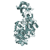
- PDB-5jbe: 4,6-alpha-glucanotransferase GTFB from Lactobacillus reuteri 121 ... -
+ Open data

- Basic information
| Entry | Database: PDB / ID: 5jbe | |||||||||
|---|---|---|---|---|---|---|---|---|---|---|
| Title | 4,6-alpha-glucanotransferase GTFB from Lactobacillus reuteri 121 complexed with an isomalto-maltopentasaccharide | |||||||||
 Components Components | Inactive glucansucrase | |||||||||
 Keywords Keywords | TRANSFERASE / 4 / 6-alpha-glucanotransferase / starch conversion / GH70 | |||||||||
| Function / homology |  Function and homology information Function and homology informationdextransucrase activity / dextransucrase / glucan biosynthetic process / glucosyltransferase activity / metal ion binding Similarity search - Function | |||||||||
| Biological species |  Lactobacillus reuteri (bacteria) Lactobacillus reuteri (bacteria) | |||||||||
| Method |  X-RAY DIFFRACTION / SYNCHROTRON / MOLECULAR REPLACEMENT / molecular replacement / Resolution: 2.1 Å X-RAY DIFFRACTION / SYNCHROTRON / MOLECULAR REPLACEMENT / molecular replacement / Resolution: 2.1 Å | |||||||||
 Authors Authors | Pijning, T. / Dijkstra, B.W. / Bai, Y. / Gangoiti-Munecas, J. / Dijkhuizen, L. | |||||||||
| Funding support |  China, China,  Netherlands, 2items Netherlands, 2items
| |||||||||
 Citation Citation | Journal: Structure / Year: 2017 Title: Crystal Structure of 4,6-alpha-Glucanotransferase Supports Diet-Driven Evolution of GH70 Enzymes from alpha-Amylases in Oral Bacteria. Authors: Bai, Y. / Gangoiti, J. / Dijkstra, B.W. / Dijkhuizen, L. / Pijning, T. #1: Journal: Journal of Agricultural and Food Chemistry / Year: 2016 Title: Lactobacillus reuteri Strains Convert Starch and Maltodextrins into Homoexopolysaccharides Using an Extracellular and Cell-Associated 4,6-alpha-Glucanotransferase Authors: Bai, Y. / Boger, M. / van der Kaaij, R.M. / Woortman, A.J.J. / Pijning, T. / van Leeuwen, S.S. / Lammerts van Bueren, A. / Dijkhuizen, L. | |||||||||
| History |
|
- Structure visualization
| Structure viewer | Molecule: MolmilJmol/JSmol |
|---|
- Downloads & links
-Download
| PDBx/mmCIF format | 5jbe.cif.gz | 674.6 KB | Display | PDBx/mmCIF format |
|---|---|---|---|---|
| PDB format | pdb5jbe.ent.gz | 553.1 KB | Display | PDB format |
| PDBx/mmJSON format | 5jbe.json.gz | Tree view | PDBx/mmJSON format | |
| Others |  Other downloads Other downloads |
-Validation report
| Arichive directory | https://data.pdbj.org/pub/pdb/validation_reports/jb/5jbeftp://data.pdbj.org/pub/pdb/validation_reports/jb/5jbe | HTTPS FTP |
|---|
-Related structure data
| Related structure data |  5jbdC 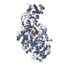 5jbfC  3klkS S: Starting model for refinement C: citing same article ( |
|---|---|
| Similar structure data |


-Links
 PDBj
PDBj


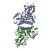
- Assembly
| Deposited unit | 
| ||||||||||||||||||
|---|---|---|---|---|---|---|---|---|---|---|---|---|---|---|---|---|---|---|---|
| 1 | 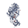
| ||||||||||||||||||
| 2 | 
| ||||||||||||||||||
| Unit cell |
| ||||||||||||||||||
| Components on special symmetry positions |
| ||||||||||||||||||
| Noncrystallographic symmetry (NCS) | NCS domain:
NCS domain segments: Component-ID: _ / Ens-ID: 1 / Beg auth comp-ID: MET / Beg label comp-ID: MET / End auth comp-ID: SER / End label comp-ID: SER / Refine code: _ / Auth seq-ID: 761 - 1614 / Label seq-ID: 1 - 854
|
-Components
-Protein , 1 types, 2 molecules AB
| #1: Protein | Mass: 94903.055 Da / Num. of mol.: 2 / Fragment: GTFB-dNdV Source method: isolated from a genetically manipulated source Source: (gene. exp.) Lactobacillus reuteri (bacteria) / Plasmid: pET15b / Production host: |
|---|
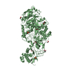
-Sugars , 3 types, 4 molecules 
| #2: Polysaccharide | Source method: isolated from a genetically manipulated source #3: Polysaccharide | alpha-D-glucopyranose-(1-4)-alpha-D-glucopyranose / alpha-maltose |   Source method: isolated from a genetically manipulated source Details: oligosaccharide / References: alpha-maltose #7: Sugar | ChemComp-BGC / |  Type: D-saccharide, beta linking / Mass: 180.156 Da / Num. of mol.: 1 Type: D-saccharide, beta linking / Mass: 180.156 Da / Num. of mol.: 1Source method: isolated from a genetically manipulated source Formula: C6H12O6 |
|---|
-Non-polymers , 4 types, 635 molecules 


| #4: Chemical |  Mass: 40.078 Da / Num. of mol.: 2 / Source method: obtained synthetically / Formula: Ca Mass: 40.078 Da / Num. of mol.: 2 / Source method: obtained synthetically / Formula: Ca#5: Chemical | ChemComp-SO4 /  Mass: 96.063 Da / Num. of mol.: 4 / Source method: obtained synthetically / Formula: SO4 Mass: 96.063 Da / Num. of mol.: 4 / Source method: obtained synthetically / Formula: SO4#6: Chemical | ChemComp-ACT /  Mass: 59.044 Da / Num. of mol.: 6 / Source method: obtained synthetically / Formula: C2H3O2 Mass: 59.044 Da / Num. of mol.: 6 / Source method: obtained synthetically / Formula: C2H3O2#8: Water | ChemComp-HOH / | Mass: 18.015 Da / Num. of mol.: 623 / Source method: isolated from a natural source / Formula: H2O |
|---|
-Experimental details
-Experiment
| Experiment | Method: X-RAY DIFFRACTION / Number of used crystals: 1 |
|---|
- Sample preparation
| Crystal | Density Matthews: 2.34 Å3/Da / Density % sol: 47.39 % |
|---|---|
| Crystal grow | Temperature: 293 K / Method: vapor diffusion, hanging drop / pH: 5 / Details: PEG3350, NaCl, (NH4)2)SO4 |
-Data collection
| Diffraction | Mean temperature: 100 K |
|---|---|
| Diffraction source | Source: SYNCHROTRON / Site: ESRF  / Beamline: ID23-2 / Wavelength: 0.8729 Å / Beamline: ID23-2 / Wavelength: 0.8729 Å |
| Detector | Type: DECTRIS PILATUS3 2M / Detector: PIXEL / Date: Oct 4, 2014 |
| Radiation | Monochromator: Si / Protocol: SINGLE WAVELENGTH / Monochromatic (M) / Laue (L): M / Scattering type: x-ray |
| Radiation wavelength | Wavelength: 0.8729 Å / Relative weight: 1 |
| Reflection | Resolution: 2.1→46.16 Å / Num. obs: 102167 / % possible obs: 99.4 % / Redundancy: 4 % / CC1/2: 0.994 / Rmerge(I) obs: 0.126 / Rpim(I) all: 0.071 / Rrim(I) all: 0.145 / Net I/σ(I): 7.7 / Num. measured all: 406237 / Scaling rejects: 3 |
| Reflection shell | Resolution: 2.1→2.14 Å / Redundancy: 3.7 % / Rmerge(I) obs: 0.818 / % possible all: 98.9 |
-Phasing
| Phasing | Method: molecular replacement | |||||||||
|---|---|---|---|---|---|---|---|---|---|---|
| Phasing MR | Model details: Phaser MODE: MR_AUTO
|
- Processing
| Software |
| |||||||||||||||||||||||||||||||||||||||||||||||||||||||||||||||||||||||||||
|---|---|---|---|---|---|---|---|---|---|---|---|---|---|---|---|---|---|---|---|---|---|---|---|---|---|---|---|---|---|---|---|---|---|---|---|---|---|---|---|---|---|---|---|---|---|---|---|---|---|---|---|---|---|---|---|---|---|---|---|---|---|---|---|---|---|---|---|---|---|---|---|---|---|---|---|---|
| Refinement | Method to determine structure: MOLECULAR REPLACEMENT Starting model: PDB ID: 3KLK domains A, B, C, IV Resolution: 2.1→46.16 Å / Cor.coef. Fo:Fc: 0.948 / Cor.coef. Fo:Fc free: 0.932 / SU B: 14.644 / SU ML: 0.185 / SU R Cruickshank DPI: 0.2655 / Cross valid method: THROUGHOUT / σ(F): 0 / ESU R: 0.262 / ESU R Free: 0.195 / Details: HYDROGENS HAVE BEEN ADDED IN THE RIDING POSITIONS
| |||||||||||||||||||||||||||||||||||||||||||||||||||||||||||||||||||||||||||
| Solvent computation | Ion probe radii: 0.9 Å / Shrinkage radii: 0.9 Å / VDW probe radii: 1.2 Å | |||||||||||||||||||||||||||||||||||||||||||||||||||||||||||||||||||||||||||
| Displacement parameters | Biso max: 90.41 Å2 / Biso mean: 39.018 Å2 / Biso min: 18.53 Å2
| |||||||||||||||||||||||||||||||||||||||||||||||||||||||||||||||||||||||||||
| Refinement step | Cycle: final / Resolution: 2.1→46.16 Å
| |||||||||||||||||||||||||||||||||||||||||||||||||||||||||||||||||||||||||||
| Refine LS restraints |
| |||||||||||||||||||||||||||||||||||||||||||||||||||||||||||||||||||||||||||
| Refine LS restraints NCS | Ens-ID: 1 / Number: 101684 / Refine-ID: X-RAY DIFFRACTION / Type: interatomic distance / Rms dev position: 0.08 Å / Weight position: 0.05
| |||||||||||||||||||||||||||||||||||||||||||||||||||||||||||||||||||||||||||
| LS refinement shell | Resolution: 2.1→2.155 Å / Total num. of bins used: 20
| |||||||||||||||||||||||||||||||||||||||||||||||||||||||||||||||||||||||||||
| Refinement TLS params. | Method: refined / Refine-ID: X-RAY DIFFRACTION
| |||||||||||||||||||||||||||||||||||||||||||||||||||||||||||||||||||||||||||
| Refinement TLS group |
|
