 Movie
Movie Controller

[English] 日本語
 Yorodumi
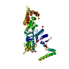
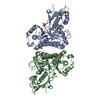
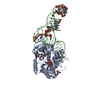
- PDB-1ls2: Fitting of EF-Tu and tRNA in the Low Resolution Cryo-EM Map of an... -
+ Open data

- Basic information
| Entry | Database: PDB / ID: 1ls2 | ||||||
|---|---|---|---|---|---|---|---|
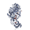
| Title | Fitting of EF-Tu and tRNA in the Low Resolution Cryo-EM Map of an EF-Tu Ternary Complex (GDP and Kirromycin) Bound to E. coli 70S Ribosome | ||||||
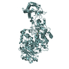
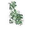
 Components Components |
| ||||||
 Keywords Keywords | TRANSLATION/RNA / EF-Tu / ternary complex / cryo-EM / 70S E.coli ribosome / TRANSLATION-RNA COMPLEX | ||||||
| Function / homology |  Function and homology information Function and homology informationguanyl-nucleotide exchange factor complex / protein-synthesizing GTPase / guanosine tetraphosphate binding / translational elongation / translation elongation factor activity / response to antibiotic / GTPase activity / GTP binding / magnesium ion binding / RNA binding ...guanyl-nucleotide exchange factor complex / protein-synthesizing GTPase / guanosine tetraphosphate binding / translational elongation / translation elongation factor activity / response to antibiotic / GTPase activity / GTP binding / magnesium ion binding / RNA binding / plasma membrane / cytoplasm Similarity search - Function | ||||||
| Biological species |   | ||||||
| Method | ELECTRON MICROSCOPY / single particle reconstruction / cryo EM / Resolution: 16.8 Å | ||||||
 Authors Authors | Valle, M. / Sengupta, J. / Swami, N.K. / Grassucci, R.A. / Burkhardt, N. / Nierhaus, K.H. / Agrawal, R.K. / Frank, J. | ||||||
 Citation Citation | Journal: EMBO J / Year: 2002 Title: Cryo-EM reveals an active role for aminoacyl-tRNA in the accommodation process. Authors: Mikel Valle / Jayati Sengupta / Neil K Swami / Robert A Grassucci / Nils Burkhardt / Knud H Nierhaus / Rajendra K Agrawal / Joachim Frank /  Abstract: During the elongation cycle of protein biosynthesis, the specific amino acid coded for by the mRNA is delivered by a complex that is comprised of the cognate aminoacyl-tRNA, elongation factor Tu and ...During the elongation cycle of protein biosynthesis, the specific amino acid coded for by the mRNA is delivered by a complex that is comprised of the cognate aminoacyl-tRNA, elongation factor Tu and GTP. As this ternary complex binds to the ribosome, the anticodon end of the tRNA reaches the decoding center in the 30S subunit. Here we present the cryo- electron microscopy (EM) study of an Escherichia coli 70S ribosome-bound ternary complex stalled with an antibiotic, kirromycin. In the cryo-EM map the anticodon arm of the tRNA presents a new conformation that appears to facilitate the initial codon-anticodon interaction. Furthermore, the elbow region of the tRNA is seen to contact the GTPase-associated center on the 50S subunit of the ribosome, suggesting an active role of the tRNA in the transmission of the signal prompting the GTP hydrolysis upon codon recognition. #1: Journal: J.Mol.Biol. / Year: 1999Title: Crystal structure of intact elongation factor EF-Tu from E.coli in GDP conformation at 2.05A resolution Authors: Song, H. / Parsons, M.R. / Rowsell, S. / Leonard, G. / Phillips, S.E. #2: Journal: Science / Year: 1995Title: Crystal structure of the ternary complex of the Phe-tRNAPhe, EF-Tu, and a GTP analog Authors: Nissen, P. / Kjeldgaard, M. / Thirup, S. / Polekhina, G. / Reshetnikova, L. / Clark, B.F.C. / Nyborg, J. #3: Journal: Nature / Year: 1997Title: Visualization of elongation factor Tu on Escherichia coli ribosome Authors: Stark, H. / Rodnina, M.V. / Rinke-Appel, J. / Brimacombe, R. / Wintermeyer, W. / van Heel, M. #4: Journal: Structure / Year: 1993Title: The crystal structure of elongation factor EF-Tu from Thermus aquaticus in the GTP conformation Authors: Kjeldgaard, M. / Nissen, P. / Thirup, S. / Nyborg, J. #5: Journal: Structure / Year: 1996Title: Helix unwinding in the effector region of elongation factor EF-Tu-GDP Authors: Polekhina, G. / Thirup, S. / Kjeldgaard, M. / Nissen, P. / Lippmann, C. / Nyborg, J. | ||||||
| History |
|
- Structure visualization
| Movie |
Movie viewer |
|---|---|
| Structure viewer | Molecule: MolmilJmol/JSmol |
 UCSF Chimera
- Downloads & links
-Download
| PDBx/mmCIF format | 1ls2.cif.gz | 30.4 KB | Display | PDBx/mmCIF format |
|---|---|---|---|---|
| PDB format | pdb1ls2.ent.gz | 13.9 KB | Display | PDB format |
| PDBx/mmJSON format | 1ls2.json.gz | Tree view | PDBx/mmJSON format | |
| Others |  Other downloads Other downloads |
-Validation report
| Arichive directory | https://data.pdbj.org/pub/pdb/validation_reports/ls/1ls2ftp://data.pdbj.org/pub/pdb/validation_reports/ls/1ls2 | HTTPS FTP |
|---|
-Related structure data
| Related structure data | 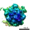 1045MC  1lu3C C: citing same article ( M: map data used to model this data |
|---|---|
| Similar structure data |


-Links
 PDBj
PDBj


- Assembly
| Deposited unit | 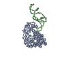
|
|---|---|
| 1 |
|
-Components
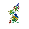
| #1: RNA chain | Mass: 24518.570 Da / Num. of mol.: 1 / Source method: isolated from a natural source / Source: (natural) |
|---|---|
| #2: Protein | Mass: 43239.297 Da / Num. of mol.: 1 / Source method: isolated from a natural source / Source: (natural) |
-Experimental details
-Experiment
| Experiment | Method: ELECTRON MICROSCOPY |
|---|---|
| EM experiment | Aggregation state: PARTICLE / 3D reconstruction method: single particle reconstruction |
- Sample preparation
| Component | Name: E. coli 70S ribosome-Ternary complex-GDP-Kirromycin / Type: RIBOSOME |
|---|---|
| Buffer solution | Name: Hepes-KOH buffer at pH 7.5 / pH: 7.5 / Details: Hepes-KOH buffer at pH 7.5 |
| Specimen | Embedding applied: NO / Shadowing applied: NO / Staining applied: NO / Vitrification applied: YES |
| Vitrification | Instrument: HOMEMADE PLUNGER / Cryogen name: ETHANE / Details: Rapid-freezing in liquid ethane |
- Electron microscopy imaging
| Microscopy | Model: FEI/PHILIPS EM420 / Date: Mar 1, 2001 |
|---|---|
| Electron gun | Electron source: LAB6 / Accelerating voltage: 100 kV / Illumination mode: FLOOD BEAM |
| Electron lens | Mode: BRIGHT FIELD / Nominal magnification: 50000 X / Calibrated magnification: 52000 X / Nominal defocus max: 3500 nm / Nominal defocus min: 1000 nm / Cs: 2 mm |
| Specimen holder | Temperature: 93 K / Tilt angle max: 0 ° / Tilt angle min: 0 ° |
| Image recording | Electron dose: 10 e/Å2 / Film or detector model: KODAK SO-163 FILM |
- Processing
| Software | Name: O / Classification: model building | |||||||||||||||||||||
|---|---|---|---|---|---|---|---|---|---|---|---|---|---|---|---|---|---|---|---|---|---|---|
| EM software |
| |||||||||||||||||||||
| CTF correction | Details: CTF correction of 3D-maps | |||||||||||||||||||||
| Symmetry | Point symmetry: C1 (asymmetric) | |||||||||||||||||||||
| 3D reconstruction | Method: reference based alignment / Resolution: 16.8 Å / Num. of particles: 7985 / Actual pixel size: 2.69 Å / Magnification calibration: TMV / Details: SPIDER package / Symmetry type: POINT | |||||||||||||||||||||
| Atomic model building | Protocol: OTHER / Space: REAL / Details: METHOD--Manual | |||||||||||||||||||||
| Atomic model building |
| |||||||||||||||||||||
| Refinement | Highest resolution: 16.8 Å | |||||||||||||||||||||
| Refinement step | Cycle: LAST / Highest resolution: 16.8 Å
|
