Movie
Movie Controller

[English] 日本語
 Yorodumi
- EMDB-1045: Cryo-EM reveals an active role for aminoacyl-tRNA in the accommod... -
+ Open data

- Basic information
| Entry | Database: EMDB / ID: EMD-1045 | |||||||||
|---|---|---|---|---|---|---|---|---|---|---|
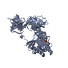
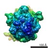
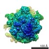
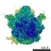
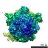
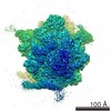
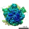
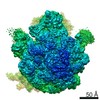
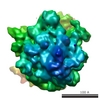
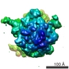
| Title | Cryo-EM reveals an active role for aminoacyl-tRNA in the accommodation process. | |||||||||
 Map data Map data | 70S aa-tRNA-EF-Tu.GDP.kirromycin complex | |||||||||
 Sample Sample |
| |||||||||
| Function / homology |  Function and homology information Function and homology informationguanyl-nucleotide exchange factor complex / protein-synthesizing GTPase / guanosine tetraphosphate binding / translational elongation / translation elongation factor activity / response to antibiotic / GTPase activity / GTP binding / magnesium ion binding / RNA binding ...guanyl-nucleotide exchange factor complex / protein-synthesizing GTPase / guanosine tetraphosphate binding / translational elongation / translation elongation factor activity / response to antibiotic / GTPase activity / GTP binding / magnesium ion binding / RNA binding / plasma membrane / cytoplasm Similarity search - Function | |||||||||
| Biological species |  | |||||||||
| Method | single particle reconstruction / cryo EM / Resolution: 16.8 Å | |||||||||
 Authors Authors | Valle M / Sengupta J / Swami NK / Grassucci RA / Burkhardt N / Nierhaus KH / Agrawal RK / Frank J | |||||||||
 Citation Citation | Journal: EMBO J / Year: 2002 Title: Cryo-EM reveals an active role for aminoacyl-tRNA in the accommodation process. Authors: Mikel Valle / Jayati Sengupta / Neil K Swami / Robert A Grassucci / Nils Burkhardt / Knud H Nierhaus / Rajendra K Agrawal / Joachim Frank /  Abstract: During the elongation cycle of protein biosynthesis, the specific amino acid coded for by the mRNA is delivered by a complex that is comprised of the cognate aminoacyl-tRNA, elongation factor Tu and ...During the elongation cycle of protein biosynthesis, the specific amino acid coded for by the mRNA is delivered by a complex that is comprised of the cognate aminoacyl-tRNA, elongation factor Tu and GTP. As this ternary complex binds to the ribosome, the anticodon end of the tRNA reaches the decoding center in the 30S subunit. Here we present the cryo- electron microscopy (EM) study of an Escherichia coli 70S ribosome-bound ternary complex stalled with an antibiotic, kirromycin. In the cryo-EM map the anticodon arm of the tRNA presents a new conformation that appears to facilitate the initial codon-anticodon interaction. Furthermore, the elbow region of the tRNA is seen to contact the GTPase-associated center on the 50S subunit of the ribosome, suggesting an active role of the tRNA in the transmission of the signal prompting the GTP hydrolysis upon codon recognition. | |||||||||
| History |
|
- Structure visualization
| Movie |
Movie viewer |
|---|---|
| Structure viewer | EM map: SurfViewMolmilJmol/JSmol |
| Supplemental images |
 UCSF Chimera
- Downloads & links
-EMDB archive
| Map data | emd_1045.map.gz | 8.8 MB | EMDB map data format | |
|---|---|---|---|---|
| Header (meta data) | emd-1045-v30.xmlemd-1045.xml | 12.6 KB 12.6 KB | Display Display | EMDB header |
| Images |  1045.gif 1045.gif | 45.1 KB | ||
| Archive directory |  http://ftp.pdbj.org/pub/emdb/structures/EMD-1045ftp://ftp.pdbj.org/pub/emdb/structures/EMD-1045 http://ftp.pdbj.org/pub/emdb/structures/EMD-1045ftp://ftp.pdbj.org/pub/emdb/structures/EMD-1045 | HTTPS FTP |
-Related structure data
| Related structure data |  1ls2MC  1lu3MC M: atomic model generated by this map C: citing same article ( |
|---|---|
| Similar structure data |

-Links
| EMDB pages | EMDB (EBI/PDBe) / EMDataResource |
|---|---|
| Related items in Molecule of the Month |


-Map
| File | Download / File: emd_1045.map.gz / Format: CCP4 / Size: 9.4 MB / Type: IMAGE STORED AS FLOATING POINT NUMBER (4 BYTES) | ||||||||||||||||||||||||||||||||||||||||||||||||||||||||||||||||||||
|---|---|---|---|---|---|---|---|---|---|---|---|---|---|---|---|---|---|---|---|---|---|---|---|---|---|---|---|---|---|---|---|---|---|---|---|---|---|---|---|---|---|---|---|---|---|---|---|---|---|---|---|---|---|---|---|---|---|---|---|---|---|---|---|---|---|---|---|---|---|
| Annotation | 70S aa-tRNA-EF-Tu.GDP.kirromycin complex | ||||||||||||||||||||||||||||||||||||||||||||||||||||||||||||||||||||
| Projections & slices | Image control
Images are generated by Spider. | ||||||||||||||||||||||||||||||||||||||||||||||||||||||||||||||||||||
| Voxel size | X=Y=Z: 2.69 Å | ||||||||||||||||||||||||||||||||||||||||||||||||||||||||||||||||||||
| Density |
| ||||||||||||||||||||||||||||||||||||||||||||||||||||||||||||||||||||
| Symmetry | Space group: 1 | ||||||||||||||||||||||||||||||||||||||||||||||||||||||||||||||||||||
| Details | EMDB XML:
CCP4 map header:
| ||||||||||||||||||||||||||||||||||||||||||||||||||||||||||||||||||||
 Z (Sec.)
 Y (Row.)
 X (Col.)


-Supplemental data
- Sample components
-Entire : 70S aa-tRNA-EF-Tu.GDP.kirromycin complex from E.coli
| Entire | Name: 70S aa-tRNA-EF-Tu.GDP.kirromycin complex from E.coli |
|---|---|
| Components |
|
-Supramolecule #1000: 70S aa-tRNA-EF-Tu.GDP.kirromycin complex from E.coli
| Supramolecule | Name: 70S aa-tRNA-EF-Tu.GDP.kirromycin complex from E.coli / type: sample / ID: 1000 / Number unique components: 6 |
|---|---|
| Molecular weight | Experimental: 2.5 MDa / Theoretical: 2.5 MDa |
-Supramolecule #1: E.coli 70S ribosome
| Supramolecule | Name: E.coli 70S ribosome / type: complex / ID: 1 / Details: 10 pmol / Recombinant expression: No / Ribosome-details: ribosome-prokaryote: ALL |
|---|---|
| Source (natural) | Organism: |
-Macromolecule #1: aa-tRNA
| Macromolecule | Name: aa-tRNA / type: rna / ID: 1 Details: 14 pmol; fMet-tRNA(fMet) at the P site and Phe-tRNA(phe) at the A site Classification: TRANSFER / Structure: DOUBLE HELIX / Synthetic?: No |
|---|---|
| Source (natural) | Organism: |
-Macromolecule #2: EF-Tu
| Macromolecule | Name: EF-Tu / type: ligand / ID: 2 / Details: 20 pmol / Recombinant expression: No |
|---|---|
| Source (natural) | Organism: |
-Macromolecule #3: GDP
| Macromolecule | Name: GDP / type: ligand / ID: 3 Details: 8 ul of 2.5mM; [Alpha-(32)P]GTP The radioactivity contributed by the Alpha-(32)P moiety of the GDP served to calculate a ~50% occupancy for the elongation factor. Recombinant expression: No |
|---|---|
| Source (natural) | Organism: synthetic construct (others) |
-Macromolecule #4: kirromycin
| Macromolecule | Name: kirromycin / type: ligand / ID: 4 / Details: 3 ul of 100 mM; antibiotic / Recombinant expression: No |
|---|---|
| Source (natural) | Organism: synthetic construct (others) |
| Chemical component information |  ChemComp-KIR: |
-Macromolecule #5: MF-mRNA
| Macromolecule | Name: MF-mRNA / type: ligand / ID: 5 / Details: 15 pmol; M(Met).F(Phe)-mRNA / Recombinant expression: No |
|---|---|
| Source (natural) | Organism: |
-Experimental details
-Structure determination
| Method | cryo EM |
|---|---|
 Processing Processing | single particle reconstruction |
| Aggregation state | particle |
-Sample preparation
| Buffer | pH: 7.5 Details: 20 mM HEPES-KOH, 6mM MgCl2, 150mM NH4Cl, 4mM 2-metcaptoethanol, 0.05 mM spermine and 2 mM spermidine |
|---|---|
| Vitrification | Cryogen name: ETHANE / Chamber humidity: 30 % / Chamber temperature: 93 K / Instrument: HOMEMADE PLUNGER Details: Vitrification instrument: two side blotting plunger Method: Blot for 2 seconds before plunging |
- Electron microscopy
| Microscope | FEI/PHILIPS EM420 |
|---|---|
| Temperature | Average: 93 K |
| Details | Microscope: Philips EM420 Imaging date: 1997 |
| Date | Jun 1, 1998 |
| Image recording | Category: FILM / Film or detector model: KODAK SO-163 FILM / Digitization - Scanner: ZEISS SCAI / Digitization - Sampling interval: 14 µm / Number real images: 45 / Average electron dose: 10 e/Å2 / Od range: 1.2 / Bits/pixel: 12 |
| Electron beam | Acceleration voltage: 100 kV / Electron source: LAB6 |
| Electron optics | Calibrated magnification: 52000 / Illumination mode: FLOOD BEAM / Imaging mode: BRIGHT FIELD / Cs: 2.0 mm / Nominal defocus max: 3.844 µm / Nominal defocus min: 1.041 µm / Nominal magnification: 49000 |
| Sample stage | Specimen holder: Cryo transfer / Specimen holder model: GATAN LIQUID NITROGEN |
-Image processing
| CTF correction | Details: defocus groups |
|---|---|
| Final reconstruction | Applied symmetry - Point group: C1 (asymmetric) / Algorithm: OTHER / Resolution.type: BY AUTHOR / Resolution: 16.8 Å / Resolution method: FSC 0.5 CUT-OFF / Software - Name: SPIDER Details: Final map was calculated from 8 CTF corrected defocus groups Number images used: 7985 |
| Final angle assignment | Details: SPIDER definition |
| Final two d classification | Number classes: 2 |
-Atomic model buiding 1
| Initial model | PDB ID: |
|---|---|
| Software | Name: manual |
| Details | Protocol: Rigid Body. The EF-Tu domains were separately fitted by manual docking using program O (pdb codes for fitted coordinates: 1LS2, 1LU3) |
| Refinement | Protocol: RIGID BODY FIT / Target criteria: cross correlation coefficient |
| Output model | PDB-1ls2: PDB-1lu3: |