 Movie
Movie Controller

[English] 日本語
 Yorodumi
- EMDB-1370: Progression of the ribosome recycling factor through the ribosome... -
+ Open data

- Basic information
| Entry | Database: EMDB / ID: EMD-1370 | |||||||||
|---|---|---|---|---|---|---|---|---|---|---|
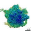
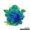
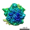
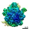
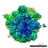
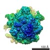
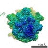
| Title | Progression of the ribosome recycling factor through the ribosome dissociates the two ribosomal subunits. | |||||||||
 Map data Map data | 70S-RRF complex | |||||||||
 Sample Sample |
| |||||||||
| Biological species |  | |||||||||
| Method | single particle reconstruction / cryo EM / Resolution: 15.3 Å | |||||||||
 Authors Authors | Barat C / Datta PP / Raj VS / Sharma MR / Kaji H / Kaji A / Agrawal RK | |||||||||
 Citation Citation | Journal: Mol Cell / Year: 2007 Title: Progression of the ribosome recycling factor through the ribosome dissociates the two ribosomal subunits. Authors: Chandana Barat / Partha P Datta / V Samuel Raj / Manjuli R Sharma / Hideko Kaji / Akira Kaji / Rajendra K Agrawal /  Abstract: After the termination step of translation, the posttermination complex (PoTC), composed of the ribosome, mRNA, and a deacylated tRNA, is processed by the concerted action of the ribosome-recycling ...After the termination step of translation, the posttermination complex (PoTC), composed of the ribosome, mRNA, and a deacylated tRNA, is processed by the concerted action of the ribosome-recycling factor (RRF), elongation factor G (EF-G), and GTP to prepare the ribosome for a fresh round of protein synthesis. However, the sequential steps of dissociation of the ribosomal subunits, and release of mRNA and deacylated tRNA from the PoTC, are unclear. Using three-dimensional cryo-electron microscopy, in conjunction with undecagold-labeled RRF, we show that RRF is capable of spontaneously moving from its initial binding site on the 70S Escherichia coli ribosome to a site exclusively on the large 50S ribosomal subunit. This movement leads to disruption of crucial intersubunit bridges and thereby to the dissociation of the two ribosomal subunits, the central event in ribosome recycling. Results of this study allow us to propose a model of ribosome recycling. | |||||||||
| History |
|
- Structure visualization
| Movie |
 Movie viewer Movie viewer |
|---|---|
| Structure viewer | EM map: SurfViewMolmilJmol/JSmol |
| Supplemental images |

- Downloads & links
-EMDB archive
| Map data | emd_1370.map.gz | 7.9 MB | EMDB map data format | |
|---|---|---|---|---|
| Header (meta data) | emd-1370-v30.xmlemd-1370.xml | 9.6 KB 9.6 KB | Display Display | EMDB header |
| Images | 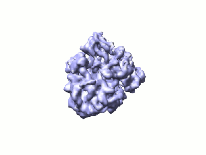 1370.gif 1370.gif | 21.6 KB | ||
| Archive directory |  http://ftp.pdbj.org/pub/emdb/structures/EMD-1370ftp://ftp.pdbj.org/pub/emdb/structures/EMD-1370 http://ftp.pdbj.org/pub/emdb/structures/EMD-1370ftp://ftp.pdbj.org/pub/emdb/structures/EMD-1370 | HTTPS FTP |
-Related structure data

-Links
| EMDB pages | EMDB (EBI/PDBe) / EMDataResource |
|---|---|
| Related items in Molecule of the Month |

-Map
| File | Download / File: emd_1370.map.gz / Format: CCP4 / Size: 8.2 MB / Type: IMAGE STORED AS FLOATING POINT NUMBER (4 BYTES) | ||||||||||||||||||||||||||||||||||||||||||||||||||||||||||||||||||||
|---|---|---|---|---|---|---|---|---|---|---|---|---|---|---|---|---|---|---|---|---|---|---|---|---|---|---|---|---|---|---|---|---|---|---|---|---|---|---|---|---|---|---|---|---|---|---|---|---|---|---|---|---|---|---|---|---|---|---|---|---|---|---|---|---|---|---|---|---|---|
| Annotation | 70S-RRF complex | ||||||||||||||||||||||||||||||||||||||||||||||||||||||||||||||||||||
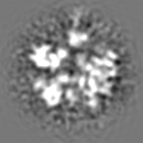
| Projections & slices | Image control
Images are generated by Spider. | ||||||||||||||||||||||||||||||||||||||||||||||||||||||||||||||||||||
| Voxel size | X=Y=Z: 2.76 Å | ||||||||||||||||||||||||||||||||||||||||||||||||||||||||||||||||||||
| Density |
| ||||||||||||||||||||||||||||||||||||||||||||||||||||||||||||||||||||
| Symmetry | Space group: 1 | ||||||||||||||||||||||||||||||||||||||||||||||||||||||||||||||||||||
| Details | EMDB XML:
CCP4 map header:
| ||||||||||||||||||||||||||||||||||||||||||||||||||||||||||||||||||||
 Z (Sec.)
 Y (Row.)
 X (Col.)


-Supplemental data
- Sample components
-Entire : 70S_RRF_UG
| Entire | Name: 70S_RRF_UG |
|---|---|
| Components |
|
-Supramolecule #1000: 70S_RRF_UG
| Supramolecule | Name: 70S_RRF_UG / type: sample / ID: 1000 / Number unique components: 2 |
|---|---|
| Molecular weight | Theoretical: 2.5205 MDa |
-Supramolecule #1: 70S Escherichia coli ribosome
| Supramolecule | Name: 70S Escherichia coli ribosome / type: complex / ID: 1 / Recombinant expression: No / Ribosome-details: ribosome-prokaryote: LSU 50S |
|---|---|
| Source (natural) | Organism: |
| Molecular weight | Experimental: 2.6 MDa |
-Macromolecule #1: Ribosome Recycling Factor
| Macromolecule | Name: Ribosome Recycling Factor / type: ligand / ID: 1 / Recombinant expression: No |
|---|---|
| Source (natural) | Organism: |
| Molecular weight | Experimental: 20.5 KDa |
-Experimental details
-Structure determination
| Method | cryo EM |
|---|---|
 Processing Processing | single particle reconstruction |
| Aggregation state | particle |
-Sample preparation
| Buffer | pH: 7.5 Details: 8.2mM MgSO4, 80mM NH4Cl, 10mM Tris (pH 7.4), without DTT |
|---|---|
| Grid | Details: Quantifoil 300 mesh Cu grid |
| Vitrification | Cryogen name: ETHANE / Chamber humidity: 100 % / Chamber temperature: 277 K / Instrument: HOMEMADE PLUNGER Details: Vitrification instrument: Wadsworth Centers fabricated guillotine plunger Method: 5ul of specimen was applied to the grid, then blotted using Whatman number 1 filter paper for 2 to 4 seconds, then plunged. |
- Electron microscopy
| Microscope | FEI TECNAI F20 |
|---|---|
| Temperature | Average: 93 K |
| Alignment procedure | Legacy - Astigmatism: objective lens astigmatism corrected and 210,000X magnification. |
| Image recording | Category: FILM / Film or detector model: KODAK SO-163 FILM / Digitization - Scanner: ZEISS SCAI / Digitization - Sampling interval: 14 µm / Number real images: 109 / Average electron dose: 20 e/Å2 / Bits/pixel: 12 |
| Electron beam | Acceleration voltage: 200 kV / Electron source:  FIELD EMISSION GUN FIELD EMISSION GUN |
| Electron optics | Calibrated magnification: 50760 / Illumination mode: FLOOD BEAM / Imaging mode: BRIGHT FIELD / Nominal defocus max: 1.8 µm / Nominal defocus min: 0.7 µm / Nominal magnification: 50000 |
| Sample stage | Specimen holder: Oxford cryo-transfer holder / Specimen holder model: OTHER |
| Experimental equipment |  Model: Tecnai F20 / Image courtesy: FEI Company |
-Image processing
| Final reconstruction | Applied symmetry - Point group: C1 (asymmetric) / Resolution.type: BY AUTHOR / Resolution: 15.3 Å / Resolution method: FSC 0.5 CUT-OFF / Software - Name: SPIDER / Number images used: 36439 |
|---|
-Atomic model buiding 1
| Software | Name: O |
|---|---|
| Details | Protocol: Rigid Body |
| Refinement | Protocol: RIGID BODY FIT |
