Movie
Movie Controller

[English] 日本語
 Yorodumi
- EMDB-1541: Visualization of the hybrid state of tRNA binding promoted by spo... -
+ Open data

- Basic information
| Entry | Database: EMDB / ID: EMD-1541 | |||||||||
|---|---|---|---|---|---|---|---|---|---|---|
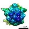
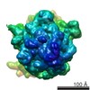
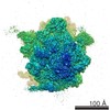
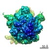
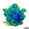
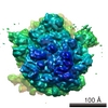
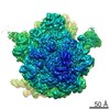
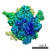
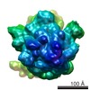
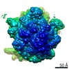
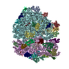
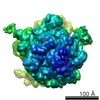
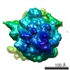
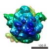
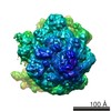
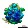
| Title | Visualization of the hybrid state of tRNA binding promoted by spontaneous ratcheting of the ribosome | |||||||||
 Map data Map data | Hybrid configuration pre-translocational ribosome | |||||||||
 Sample Sample |
| |||||||||
 Keywords Keywords | Hybrid state tRNA configuration / pre-translocational ribosome / spontaneous ratchet motion | |||||||||
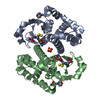
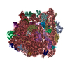
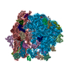
| Biological species |  | |||||||||
| Method | single particle reconstruction / cryo EM / negative staining / Resolution: 8.9 Å | |||||||||
 Authors Authors | Agirrezabala X / Lei J / Brunelle JL / Ortiz-Meoz RF / Green R / Frank J | |||||||||
 Citation Citation | Journal: Mol Cell / Year: 2008 Title: Visualization of the hybrid state of tRNA binding promoted by spontaneous ratcheting of the ribosome. Authors: Xabier Agirrezabala / Jianlin Lei / Julie L Brunelle / Rodrigo F Ortiz-Meoz / Rachel Green / Joachim Frank /  Abstract: A crucial step in translation is the translocation of tRNAs through the ribosome. In the transition from one canonical site to the other, the tRNAs acquire intermediate configurations, so-called ...A crucial step in translation is the translocation of tRNAs through the ribosome. In the transition from one canonical site to the other, the tRNAs acquire intermediate configurations, so-called hybrid states. At this stage, the small subunit is rotated with respect to the large subunit, and the anticodon stem loops reside in the A and P sites of the small subunit, while the acceptor ends interact with the P and E sites of the large subunit. In this work, by means of cryo-EM and particle classification procedures, we visualize the hybrid state of both A/P and P/E tRNAs in an authentic factor-free ribosome complex during translocation. In addition, we show how the repositioning of the tRNAs goes hand in hand with the change in the interplay between S13, L1 stalk, L5, H68, H69, and H38 that is caused by the ratcheting of the small subunit. | |||||||||
| History |
|
- Structure visualization
| Movie |
 Movie viewer Movie viewer |
|---|---|
| Structure viewer | EM map: SurfViewMolmilJmol/JSmol |
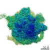
| Supplemental images |

- Downloads & links
-EMDB archive
| Map data | emd_1541.map.gz | 54.6 MB | EMDB map data format | |
|---|---|---|---|---|
| Header (meta data) | emd-1541-v30.xmlemd-1541.xml | 11.1 KB 11.1 KB | Display Display | EMDB header |
| Images |  1541.gif 1541.gif | 1.3 MB | ||
| Archive directory |  http://ftp.pdbj.org/pub/emdb/structures/EMD-1541ftp://ftp.pdbj.org/pub/emdb/structures/EMD-1541 http://ftp.pdbj.org/pub/emdb/structures/EMD-1541ftp://ftp.pdbj.org/pub/emdb/structures/EMD-1541 | HTTPS FTP |
-Related structure data


-Links
| EMDB pages | EMDB (EBI/PDBe) / EMDataResource |
|---|---|
| Related items in Molecule of the Month |

-Map
| File | Download / File: emd_1541.map.gz / Format: CCP4 / Size: 58.2 MB / Type: IMAGE STORED AS FLOATING POINT NUMBER (4 BYTES) | ||||||||||||||||||||||||||||||||||||||||||||||||||||||||||||||||||||
|---|---|---|---|---|---|---|---|---|---|---|---|---|---|---|---|---|---|---|---|---|---|---|---|---|---|---|---|---|---|---|---|---|---|---|---|---|---|---|---|---|---|---|---|---|---|---|---|---|---|---|---|---|---|---|---|---|---|---|---|---|---|---|---|---|---|---|---|---|---|
| Annotation | Hybrid configuration pre-translocational ribosome | ||||||||||||||||||||||||||||||||||||||||||||||||||||||||||||||||||||
| Projections & slices | Image control
Images are generated by Spider. | ||||||||||||||||||||||||||||||||||||||||||||||||||||||||||||||||||||
| Voxel size | X=Y=Z: 1.5 Å | ||||||||||||||||||||||||||||||||||||||||||||||||||||||||||||||||||||
| Density |
| ||||||||||||||||||||||||||||||||||||||||||||||||||||||||||||||||||||
| Symmetry | Space group: 1 | ||||||||||||||||||||||||||||||||||||||||||||||||||||||||||||||||||||
| Details | EMDB XML:
CCP4 map header:
| ||||||||||||||||||||||||||||||||||||||||||||||||||||||||||||||||||||
 Z (Sec.)
 Y (Row.)
 X (Col.)


-Supplemental data
- Sample components
-Entire : Hybrid state pre-translocational ribosome
| Entire | Name: Hybrid state pre-translocational ribosome |
|---|---|
| Components |
|
-Supramolecule #1000: Hybrid state pre-translocational ribosome
| Supramolecule | Name: Hybrid state pre-translocational ribosome / type: sample / ID: 1000 / Details: HiFi buffer, 3.5 mM Mg2 / Oligomeric state: Monomeric / Number unique components: 3 |
|---|---|
| Molecular weight | Experimental: 2.6 MDa / Theoretical: 2.6 MDa / Method: Sedimentation |
-Supramolecule #1: 70S prokaryote ribosome
| Supramolecule | Name: 70S prokaryote ribosome / type: complex / ID: 1 / Name.synonym: Ribosome / Details: Pre-translocational ribosome / Recombinant expression: No / Ribosome-details: ribosome-prokaryote: ALL |
|---|---|
| Source (natural) | Organism: |
| Molecular weight | Experimental: 2.6 MDa / Theoretical: 2.6 MDa |
-Experimental details
-Structure determination
| Method | negative staining, cryo EM |
|---|---|
 Processing Processing | single particle reconstruction |
| Aggregation state | particle |
-Sample preparation
| Concentration | 0.07 mg/mL |
|---|---|
| Buffer | pH: 7.5 Details: HiFi (50 mM Tris-HCl pH 7.5, 70mM NH4Cl, 30 mM KCl, 3.5 mM MgCl2, 0.5 mM spermidine, 8mM putrescine, 2 mM DTT) |
| Staining | Type: NEGATIVE / Details: Cryo-sample |
| Grid | Details: Quantifoil 2/4 |
| Vitrification | Cryogen name: NITROGEN / Chamber humidity: 90 % / Chamber temperature: 80 K / Instrument: OTHER / Details: Vitrification instrument: Vitrobot / Method: Blot for 6 seconds before plunging |
- Electron microscopy
| Microscope | FEI TECNAI F30 |
|---|---|
| Temperature | Min: 80.7 K / Max: 80.7 K / Average: 80.7 K |
| Alignment procedure | Legacy - Astigmatism: Objective corrected at 100,000 times magnification |
| Specialist optics | Energy filter - Name: No energy filter |
| Details | Automated data collection system AutoEMation (CCD mag. 100000x) |
| Date | Apr 15, 2007 |
| Image recording | Category: CCD / Film or detector model: TVIPS TEMCAM-F415 (4k x 4k) / Average electron dose: 25 e/Å2 Details: Automated data collection system AutoEMation (CCD mag. 100000x) TVIPS TemCam-F415 |
| Tilt angle min | 0 |
| Tilt angle max | 0 |
| Electron beam | Acceleration voltage: 300 kV / Electron source:  FIELD EMISSION GUN FIELD EMISSION GUN |
| Electron optics | Calibrated magnification: 58269 / Illumination mode: FLOOD BEAM / Imaging mode: BRIGHT FIELD / Cs: 2.26 mm / Nominal defocus max: 4.0 µm / Nominal defocus min: 1.2 µm / Nominal magnification: 59000 |
| Sample stage | Specimen holder: Cartridge / Specimen holder model: OTHER |
| Experimental equipment |  Model: Tecnai F30 / Image courtesy: FEI Company |